
Transapical heart valve delivery system and method
a delivery system and transapical heart valve technology, applied in balloon catheters, medical science, surgery, etc., can solve the problems of high invasiveness of valve replacement surgery, adverse reactions, bleeding, sudden death, etc., and achieve the effect of facilitating the positioning of the valv
Patent Information
- Authority / Receiving Office
- US · United States
- Patent Type
- Patents(United States)
- Current Assignee / Owner
- Publication Date
- 2014-07-01
Smart Images
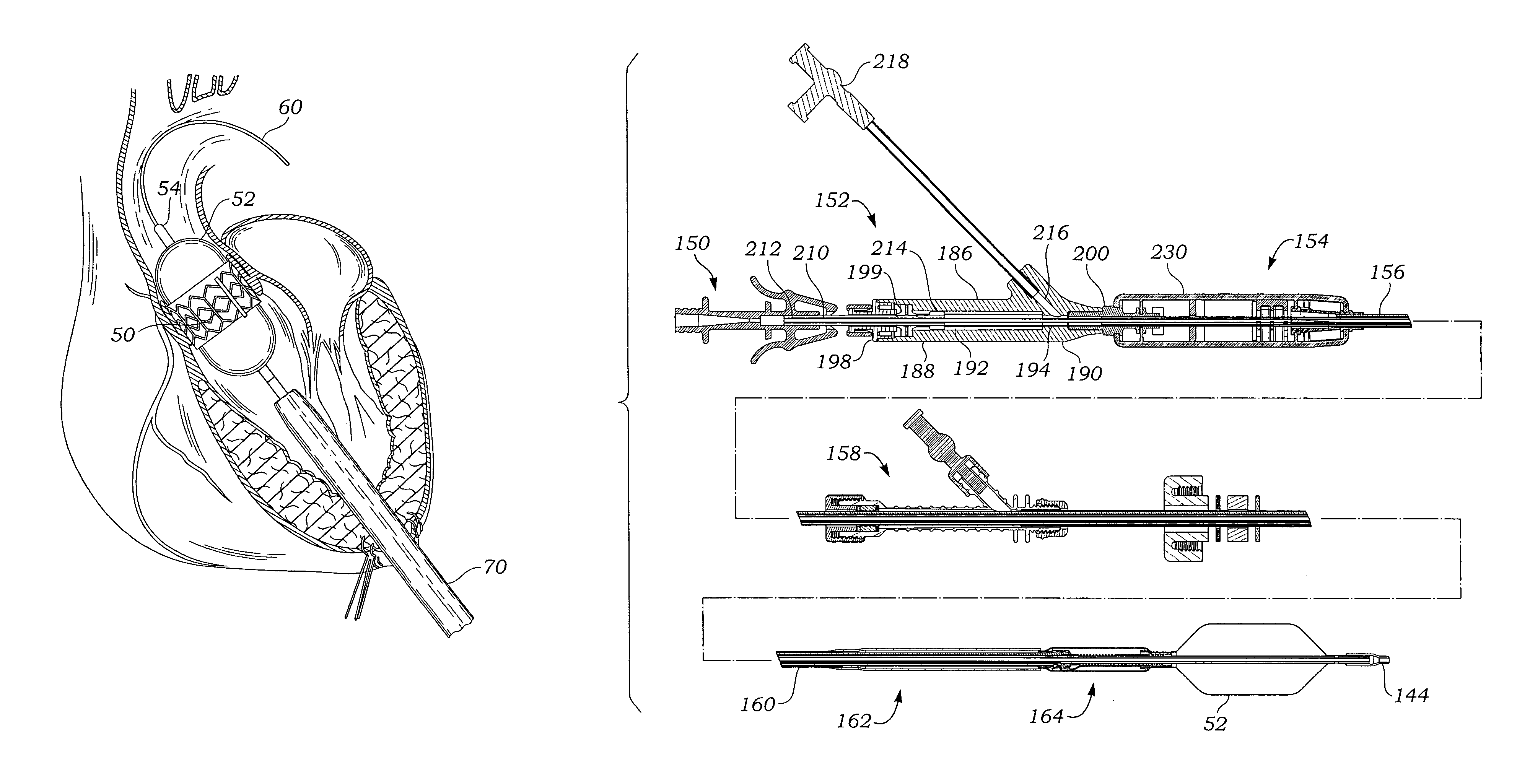
Figure 1 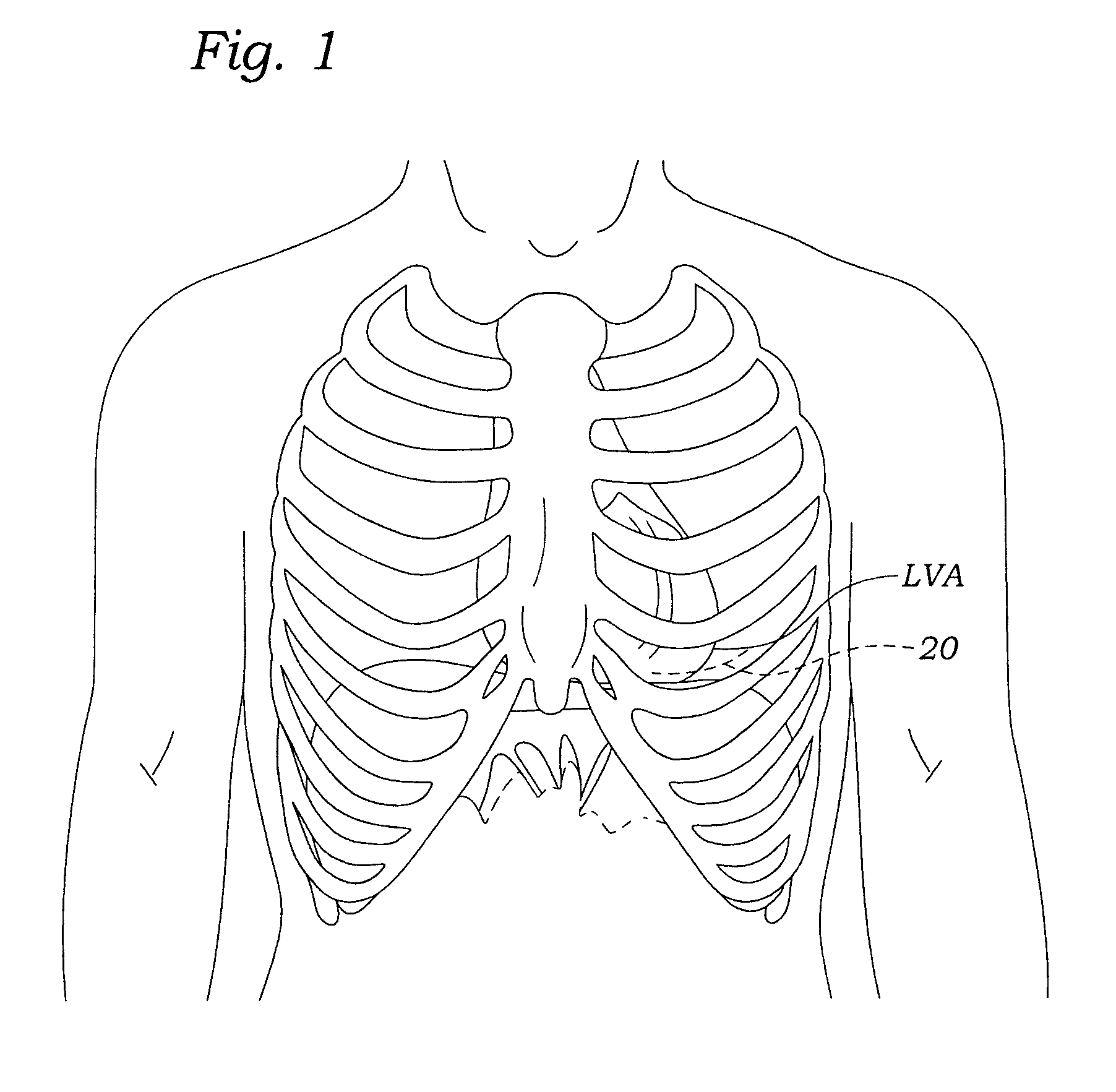
Figure 2 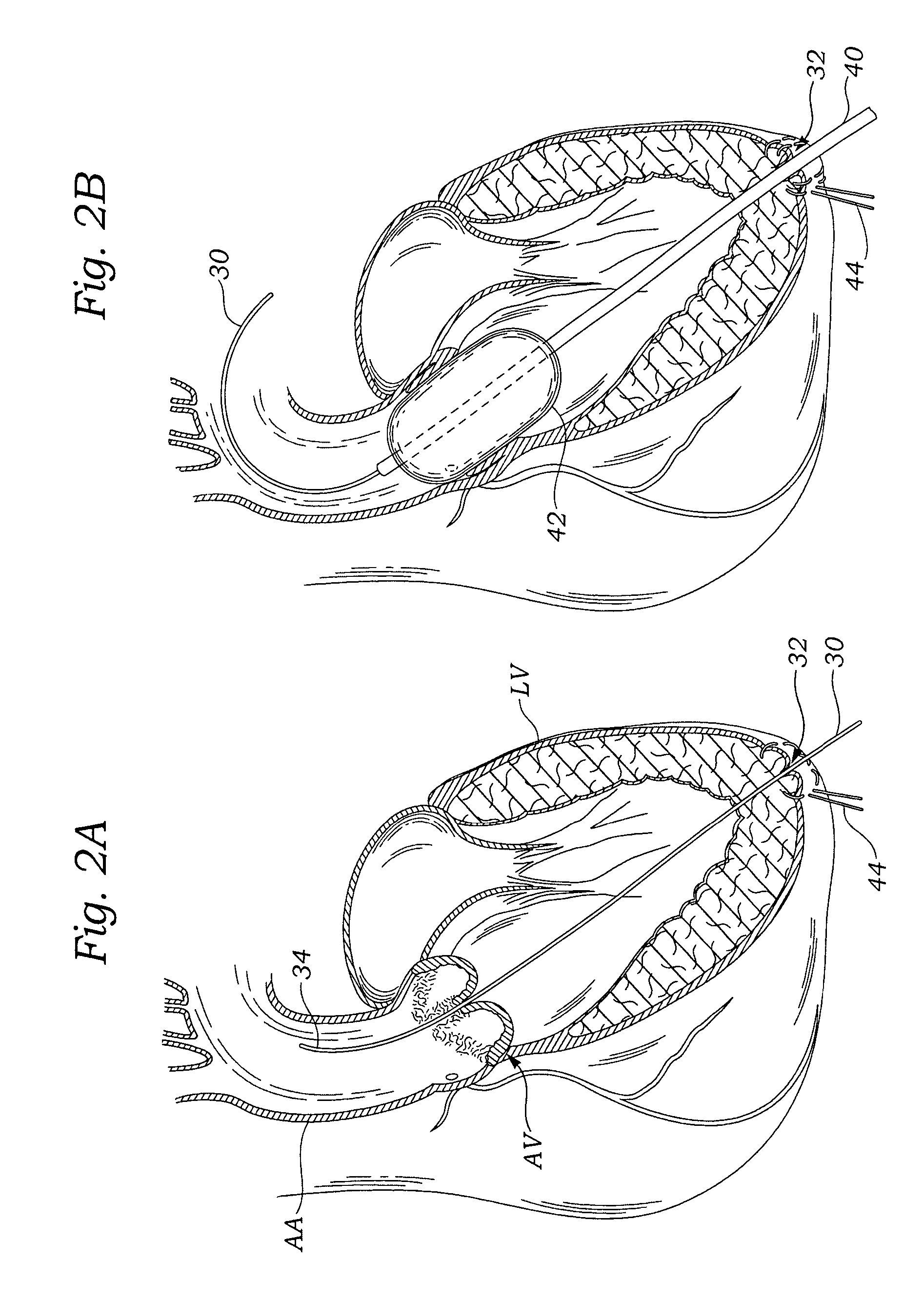
Figure 3
Abstract
Description
FIELD OF THE INVENTION
[0001] The present invention relates to methods and systems used to deliver a prosthetic valve to a heart. More specifically, the present invention relates to methods and apparatus for surgically replacing a heart valve without opening the chest cavity and with or without placing the patient on bypass, the latter being termed “off-pump.”BACKGROUND OF THE INVENTION
[0002] Heart valve replacement may be indicated when there is a narrowing of the native heart valve, commonly referred to as stenosis, or when the native valve leaks or regurgitates, such as when the leaflets are calcified. When replacing the valve, the native valve may be excised and replaced with either a biologic or a mechanical valve. Mechanical valves require lifelong anticoagulant medication to prevent blood clot formation, and clicking of the valve often may be heard through the chest. Biologic tissue valves typically do not require such medication. Tissue valves may be obtained from cadavers or m...
Examples
Embodiment Construction
[0040]The heart is a hollow muscular organ of a somewhat conical form; it lies between the lungs in the middle mediastinum and is enclosed in the pericardium. The heart rests obliquely in the chest behind the body of the sternum and adjoining parts of the rib cartilages, and projects farther into the left than into the right half of the thoracic cavity so that about one-third is situated on the right and two-thirds on the left of the median plane. The heart is subdivided by septa into right and left halves, and a constriction subdivides each half of the organ into two cavities, the upper cavity being called the atrium, the lower the ventricle. The heart therefore consists of four chambers; the right and left atria, and right and left ventricles.
[0041]As seen in FIG. 1, the left ventricular apex LVA is directed downward, forward, and to the left (from the perspective of the patient). The apex typically lies behind the fifth left intercostal space (or between the fourth and fifth), 8 ...