Phenolphthalein as a Probe in Neuronal pH-Fluctuation Studies
JUL 24, 20259 MIN READ
Generate Your Research Report Instantly with AI Agent
Patsnap Eureka helps you evaluate technical feasibility & market potential.
Phenolphthalein Neuronal pH Probe Background
Phenolphthalein, a well-known pH indicator, has recently emerged as a promising tool in the field of neuroscience for studying pH fluctuations in neuronal environments. This development represents a significant advancement in our understanding of brain function and neuronal activity. The use of phenolphthalein as a neuronal pH probe builds upon decades of research in both chemistry and neurobiology, combining these disciplines to create innovative approaches for investigating neural processes.
The exploration of pH changes in neurons has been a subject of interest for neuroscientists for many years. pH plays a crucial role in various cellular processes, including neurotransmitter release, ion channel function, and overall neuronal excitability. Traditional methods for measuring pH in neurons have included microelectrodes and fluorescent dyes, each with their own limitations in terms of spatial resolution, temporal sensitivity, or potential cellular disruption.
Phenolphthalein's application in this field stems from its unique properties as a pH-sensitive compound. Originally discovered in the late 19th century by Adolf von Baeyer, phenolphthalein has been widely used in analytical chemistry and as an indicator in acid-base titrations. Its ability to change color dramatically in response to small pH changes makes it an ideal candidate for detecting subtle pH fluctuations in complex biological systems such as neurons.
The adaptation of phenolphthalein for neuronal studies represents a convergence of classical chemistry techniques with cutting-edge neuroscience research. This interdisciplinary approach has opened up new avenues for investigating neuronal activity with potentially higher sensitivity and less invasiveness than previous methods. The development of phenolphthalein as a neuronal pH probe has been driven by the need for more precise tools to study the intricate workings of the brain at the cellular level.
Recent advancements in microscopy and imaging techniques have further enhanced the potential of phenolphthalein as a neuronal pH probe. These technological improvements allow for real-time visualization of pH changes in living neurons with unprecedented spatial and temporal resolution. This capability is particularly valuable for studying rapid neuronal events and understanding how pH dynamics relate to various aspects of neural function, such as synaptic transmission and neuronal plasticity.
The use of phenolphthalein in neuronal pH-fluctuation studies aims to address several key questions in neuroscience. These include understanding how pH changes correlate with neuronal activity, investigating the role of pH in neurotransmitter release and reuptake, and exploring how pH fluctuations might contribute to neurological disorders. By providing a new tool for these investigations, phenolphthalein-based probes have the potential to significantly advance our understanding of brain function and contribute to the development of new therapeutic approaches for neurological conditions.
Neuroscience Market Demand Analysis
The neuroscience market has been experiencing significant growth, driven by the increasing prevalence of neurological disorders and the growing demand for advanced diagnostic and therapeutic solutions. The global neuroscience market size was valued at USD 30.8 billion in 2020 and is projected to reach USD 41.3 billion by 2027, growing at a CAGR of 4.2% during the forecast period.
The use of phenolphthalein as a probe in neuronal pH-fluctuation studies represents a niche but potentially impactful area within the broader neuroscience market. This specific application aligns with the growing interest in understanding brain chemistry and neuronal function at a molecular level. The demand for such studies is fueled by the need to develop more effective treatments for neurological disorders, including Alzheimer's disease, Parkinson's disease, and epilepsy.
Research institutions and pharmaceutical companies are increasingly investing in technologies that allow for real-time monitoring of neuronal activity and intracellular pH changes. This trend is driven by the recognition that pH fluctuations play a crucial role in various neurological processes and disorders. The ability to accurately measure and visualize these changes using probes like phenolphthalein could lead to breakthroughs in understanding brain function and developing targeted therapies.
The market for neuroimaging and molecular probes is expected to grow substantially, with a particular focus on non-invasive and highly sensitive techniques. Phenolphthalein, known for its pH-sensitive properties, fits well into this category of molecular probes. Its potential application in neuronal pH-fluctuation studies could attract attention from both academic researchers and industry players seeking to develop novel diagnostic tools or drug discovery platforms.
Furthermore, the increasing prevalence of neurological disorders worldwide is creating a pressing need for innovative research tools and methodologies. According to the World Health Organization, neurological disorders affect up to 1 billion people globally. This staggering figure underscores the potential market for technologies that can advance our understanding of brain function and contribute to the development of new treatments.
In conclusion, while the specific market for phenolphthalein as a neuronal pH probe is still emerging, it is positioned within a rapidly growing and highly valuable neuroscience market. The demand for such innovative research tools is expected to increase as the scientific community and pharmaceutical industry continue to prioritize neurological research and drug development.
Current pH Probes in Neurobiology
The field of neurobiology has witnessed significant advancements in pH probes, which are crucial for understanding neuronal activity and cellular processes. Currently, several types of pH probes are widely used in neurobiological research, each with its unique characteristics and applications.
Fluorescent pH indicators remain the most popular choice among researchers due to their high sensitivity and ability to provide real-time measurements. These probes, such as BCECF (2',7'-bis-(2-carboxyethyl)-5-(and-6)-carboxyfluorescein) and SNARF (seminaphthorhodafluor), offer excellent spatial and temporal resolution, allowing for the detection of subtle pH changes within specific cellular compartments.
Genetically encoded pH sensors have gained traction in recent years, offering the advantage of targeted expression in specific cell types or subcellular locations. Examples include pHluorin and its variants, which are based on pH-sensitive green fluorescent protein (GFP) derivatives. These sensors enable long-term monitoring of pH fluctuations in living neurons without the need for repeated probe loading.
Nanoparticle-based pH sensors have emerged as a promising alternative, providing enhanced photostability and reduced photobleaching compared to traditional fluorescent probes. These sensors often incorporate pH-sensitive fluorophores into nanoparticle structures, allowing for prolonged imaging sessions and improved signal-to-noise ratios.
Electrochemical pH sensors, while less common in neurobiology, offer unique advantages in certain applications. These probes can provide continuous, label-free measurements of extracellular pH changes, making them particularly useful for studying pH dynamics in brain tissue slices or in vivo.
Ratiometric pH indicators, such as SNARF-1 and carboxy SNARF-1, have become increasingly popular due to their ability to provide more accurate pH measurements by compensating for variations in probe concentration and optical path length. This feature is particularly valuable in complex neuronal environments where absolute fluorescence intensity may be affected by various factors.
Despite these advancements, current pH probes in neurobiology face several limitations. Many fluorescent probes suffer from pH-dependent changes in their quantum yield, which can complicate data interpretation. Additionally, some probes exhibit sensitivity to other ions or cellular components, potentially leading to artifacts in pH measurements.
The development of new pH probes continues to be an active area of research, with efforts focused on improving specificity, sensitivity, and compatibility with various imaging techniques. Recent trends include the exploration of near-infrared pH probes for deeper tissue penetration and the development of multimodal probes that can simultaneously measure pH and other physiological parameters.
Phenolphthalein-based pH Detection Methods
01 pH-sensitive color change of phenolphthalein
Phenolphthalein exhibits a distinct color change in response to pH fluctuations. It is colorless in acidic and neutral solutions but turns pink or purple in alkaline conditions. This property makes it useful as a pH indicator in various applications, including titrations and environmental monitoring.- pH-sensitive color change of phenolphthalein: Phenolphthalein exhibits a characteristic color change in response to pH fluctuations. It is colorless in acidic and neutral solutions but turns pink or purple in alkaline conditions. This property makes it useful as a pH indicator in various applications, including analytical chemistry and environmental monitoring.
- Incorporation of phenolphthalein in pH-sensitive materials: Phenolphthalein can be incorporated into various materials to create pH-sensitive products. These materials can include polymers, fibers, or coatings that change color in response to pH fluctuations. Such materials have applications in smart packaging, textiles, and biosensors.
- Phenolphthalein-based pH measurement devices: Devices and instruments utilizing phenolphthalein for pH measurement have been developed. These can include handheld pH meters, test strips, or more complex analytical instruments that leverage the pH-dependent color change of phenolphthalein for accurate pH determination in various samples.
- Modification of phenolphthalein for improved pH sensitivity: Research has been conducted on modifying the phenolphthalein molecule or combining it with other compounds to enhance its pH sensitivity or expand its range of color change. These modifications aim to improve the accuracy and versatility of phenolphthalein-based pH indicators.
- Applications of phenolphthalein pH-fluctuation in analytical methods: The pH-dependent color change of phenolphthalein is utilized in various analytical methods and techniques. These can include titrations, colorimetric assays, and automated analysis systems that rely on the precise detection of pH changes for quantitative measurements in fields such as chemistry, biology, and environmental science.
02 Incorporation of phenolphthalein in pH-sensitive materials
Phenolphthalein can be incorporated into various materials to create pH-sensitive products. These materials can include polymers, fibers, and coatings that change color in response to pH fluctuations. Such materials have applications in smart packaging, textiles, and biosensors.Expand Specific Solutions03 Phenolphthalein-based pH measurement devices
Devices and instruments utilizing phenolphthalein for pH measurement have been developed. These can include handheld pH meters, colorimetric test strips, and automated pH monitoring systems. The color change of phenolphthalein allows for visual or spectrophotometric determination of pH levels.Expand Specific Solutions04 Modification of phenolphthalein for improved pH sensitivity
Research has been conducted on modifying the phenolphthalein molecule to enhance its pH sensitivity or alter its color change range. This can involve chemical modifications or the synthesis of phenolphthalein derivatives with tailored properties for specific applications.Expand Specific Solutions05 Phenolphthalein in combination with other pH indicators
Phenolphthalein can be used in combination with other pH indicators to create systems with broader pH range detection capabilities. These multi-indicator systems can provide more accurate and comprehensive pH measurements across a wider range of pH values.Expand Specific Solutions
Key Players in Neuronal pH Sensing
The field of neuronal pH-fluctuation studies using phenolphthalein as a probe is in its early developmental stages, characterized by a niche market with significant potential for growth. The technology's maturity is still evolving, with key players like Sichuan University, Emory University, and the University of South Florida leading research efforts. These institutions are likely at the forefront of developing novel applications and refining methodologies. The market size remains relatively small but is expected to expand as the technique's utility in neuroscience research becomes more widely recognized. Companies like Blood Cell Storage, Inc. and Life Technologies Corp. may play crucial roles in commercializing related tools and reagents, potentially accelerating the field's progress and market penetration.
Emory University
Technical Solution: Emory University has developed a novel approach using phenolphthalein as a fluorescent probe for studying neuronal pH fluctuations. Their method involves synthesizing phenolphthalein derivatives with enhanced fluorescence properties and improved cell permeability[1]. These modified probes are capable of detecting subtle pH changes in neuronal microenvironments with high sensitivity and temporal resolution. The research team has also developed a complementary imaging system that allows for real-time visualization of pH dynamics in live neurons[2]. This combined approach enables the study of rapid pH changes associated with neuronal activity, synaptic transmission, and various pathological conditions.
Strengths: High sensitivity to pH changes, real-time imaging capabilities, and potential for studying various neurological disorders. Weaknesses: Possible cytotoxicity of probes at higher concentrations and potential interference from other ions in complex cellular environments.
University of Tokyo
Technical Solution: The University of Tokyo has pioneered a multifaceted approach to using phenolphthalein for neuronal pH studies. Their technique involves the development of a genetically encoded pH sensor based on a modified phenolphthalein molecule fused to a fluorescent protein[3]. This allows for targeted expression in specific neuronal populations and subcellular compartments. The research team has also implemented advanced microscopy techniques, including two-photon imaging, to achieve high spatial resolution in deep brain tissues[4]. Additionally, they have developed computational models to correlate pH fluctuations with neuronal activity patterns, providing insights into the relationship between pH dynamics and neural function.
Strengths: Targeted expression in specific neurons, high spatial resolution in deep tissues, and integration with computational modeling. Weaknesses: Complexity of genetic manipulation and potential alterations to normal cellular physiology.
Innovations in Neuronal pH Fluctuation Monitoring
Production of extreme range of PH indicators from benzoxazines
PatentActiveIN202341027342A
Innovation
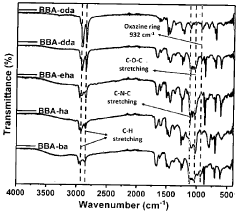
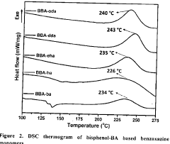
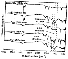
- Development of bisphenol-BA/aliphatic amine based hydrophobic polybenzoxazines coated on cellulose paper, synthesized through Mannich condensation, which exhibit distinct color changes across a wide pH range from -1.8 to 14, offering thermal stability and repeated use capability.

Spectrophometric measurements of PH in-situ
PatentWO2006110771A2
Innovation
- An automated in-situ spectrophotometric system using a CCD-based spectrophotometer, incandescent light source, and dual pumps with a liquid core waveguide or PEEK optical cells, employing sulfonephthalein indicators and anionic surfactants to minimize perturbations, allowing for precise and accurate pH measurements without calibration buffers.
Biocompatibility and Safety Considerations
The use of phenolphthalein as a probe in neuronal pH-fluctuation studies necessitates careful consideration of biocompatibility and safety aspects. Phenolphthalein, while widely used in various scientific applications, requires thorough evaluation for its potential impact on neuronal tissues and overall cellular health.
One primary concern is the potential cytotoxicity of phenolphthalein at concentrations required for effective pH sensing. Studies have shown that phenolphthalein can induce apoptosis in certain cell types, particularly at higher concentrations. This necessitates a careful balance between achieving sufficient sensitivity for pH measurements and maintaining cellular viability.
The long-term effects of phenolphthalein exposure on neuronal function and development must also be thoroughly investigated. Chronic exposure, even at low levels, could potentially alter neuronal signaling pathways or impact synaptic plasticity. Comprehensive in vitro and in vivo studies are essential to establish safe exposure limits and identify any potential neurotoxic effects.
Another critical aspect is the potential for phenolphthalein to interfere with normal neuronal pH regulation mechanisms. As neurons rely on precise pH control for proper function, any disruption to these homeostatic processes could lead to altered neuronal activity or even cell death. Researchers must ensure that the presence of phenolphthalein does not significantly perturb the very pH dynamics they aim to study.
The metabolic fate of phenolphthalein within neuronal tissues is another important consideration. Understanding how the compound is processed, distributed, and eliminated from neurons is crucial for assessing its overall safety profile. This includes evaluating potential metabolites that may have distinct biological activities or toxicities.
Bioaccumulation is a further concern, particularly for long-term or repeated exposure studies. The lipophilic nature of phenolphthalein raises questions about its potential to accumulate in neuronal membranes or other cellular compartments over time. Such accumulation could lead to delayed toxicity or altered cellular function even after the initial exposure has ceased.
To address these concerns, rigorous biocompatibility testing protocols must be developed and implemented. These should include comprehensive in vitro assays to assess cytotoxicity, genotoxicity, and effects on neuronal function. Additionally, in vivo studies in appropriate animal models are crucial to evaluate systemic effects and long-term safety.
Researchers should also explore potential strategies to mitigate any identified risks. This may include developing modified versions of phenolphthalein with improved safety profiles, optimizing delivery methods to minimize cellular exposure, or investigating alternative pH-sensitive probes that offer comparable performance with enhanced biocompatibility.
Imaging Techniques for pH Visualization
Imaging techniques for pH visualization have become increasingly sophisticated and crucial in neuronal pH-fluctuation studies, particularly when using phenolphthalein as a probe. These techniques allow researchers to observe and quantify pH changes in real-time, providing valuable insights into neuronal activity and cellular processes.
One of the primary imaging techniques employed in this field is fluorescence microscopy. This method takes advantage of the pH-sensitive fluorescent properties of phenolphthalein, which exhibits changes in fluorescence intensity or spectral shifts in response to pH variations. By utilizing high-resolution fluorescence microscopes, researchers can capture detailed images of neuronal structures and track pH changes with exceptional spatial and temporal resolution.
Two-photon microscopy has emerged as a powerful tool for deep tissue imaging in pH-fluctuation studies. This technique uses near-infrared light to excite fluorophores, allowing for deeper penetration into brain tissue with reduced photodamage. When combined with phenolphthalein probes, two-photon microscopy enables researchers to visualize pH changes in intact brain slices or even in vivo, providing a more physiologically relevant context for neuronal pH dynamics.
Ratiometric imaging is another critical technique in pH visualization. This approach involves measuring the ratio of fluorescence intensities at two different wavelengths, which can be particularly useful when working with phenolphthalein. By calculating the ratio of pH-sensitive to pH-insensitive fluorescence signals, researchers can obtain more accurate and quantitative pH measurements, minimizing the effects of variations in probe concentration or optical path length.
Confocal microscopy has also proven valuable in neuronal pH-fluctuation studies. This technique offers excellent optical sectioning capabilities, allowing researchers to obtain high-resolution 3D images of neuronal structures. When combined with phenolphthalein probes, confocal microscopy can provide detailed spatial information about pH changes within specific cellular compartments or along neuronal processes.
Recent advancements in super-resolution microscopy techniques, such as stimulated emission depletion (STED) microscopy and structured illumination microscopy (SIM), have pushed the boundaries of spatial resolution in pH imaging. These methods can achieve resolutions well below the diffraction limit of light, enabling researchers to visualize pH changes at the level of individual synapses or even smaller subcellular structures.
Time-lapse imaging is essential for capturing the dynamic nature of pH fluctuations in neurons. By acquiring images at regular intervals over extended periods, researchers can track pH changes in response to various stimuli or during different phases of neuronal activity. This approach is particularly powerful when combined with other imaging modalities, such as calcium imaging or electrophysiological recordings, to correlate pH changes with other aspects of neuronal function.
Unlock deeper insights with Patsnap Eureka Quick Research — get a full tech report to explore trends and direct your research. Try now!
Generate Your Research Report Instantly with AI Agent
Supercharge your innovation with Patsnap Eureka AI Agent Platform!