Select Atomic Force Microscopy Mode For Precise Imaging — Parameters
SEP 19, 20259 MIN READ
Generate Your Research Report Instantly with AI Agent
Patsnap Eureka helps you evaluate technical feasibility & market potential.
AFM Technology Background and Objectives
Atomic Force Microscopy (AFM) has evolved significantly since its invention in 1986 by Gerd Binnig, Calvin Quate, and Christoph Gerber. This revolutionary scanning probe microscopy technique has transformed our ability to visualize and manipulate matter at the nanoscale, offering unprecedented resolution down to the atomic level. Unlike electron microscopy, AFM does not require vacuum conditions or sample conductivity, making it versatile across various scientific disciplines including materials science, biology, and semiconductor research.
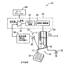
The fundamental principle of AFM involves a cantilever with a sharp tip that interacts with the sample surface. As the tip scans across the surface, various forces between the tip and sample cause deflections of the cantilever, which are measured using optical detection systems. These measurements create topographical maps with remarkable precision, often achieving sub-nanometer resolution in the vertical dimension.
Over the decades, AFM technology has branched into multiple specialized modes, each designed to extract specific information from samples. Contact mode, the original implementation, has been supplemented by tapping mode, non-contact mode, and more advanced techniques such as force spectroscopy, conductive AFM, and magnetic force microscopy. This diversification reflects the growing sophistication of research needs across scientific domains.
The primary objective in AFM imaging is achieving optimal resolution while minimizing sample damage. This delicate balance requires careful parameter selection based on sample characteristics, environmental conditions, and the specific information sought. Parameters such as cantilever stiffness, resonance frequency, scan rate, feedback gains, and setpoint values critically influence image quality and data reliability.
Recent technological advancements have focused on enhancing imaging speed, improving force sensitivity, and developing multimodal capabilities that simultaneously capture complementary data channels. High-speed AFM now enables visualization of dynamic biological processes in real-time, while ultra-sensitive force detection allows for mapping of subtle molecular interactions.
The current frontier in AFM development aims to standardize parameter selection protocols for specific applications, automate optimization processes, and integrate machine learning algorithms for intelligent scanning and data interpretation. These efforts address the persistent challenge of reproducibility in AFM measurements and seek to make this powerful technique more accessible to researchers without extensive technical expertise.
As nanoscience and nanotechnology continue to advance, precise AFM imaging becomes increasingly crucial for characterizing novel materials, understanding biological systems, and developing next-generation devices. The ability to select appropriate AFM modes and optimize scanning parameters represents a key competency for researchers seeking to extract meaningful, reliable data from this versatile microscopy platform.
Market Applications and Demand Analysis
The Atomic Force Microscopy (AFM) market has experienced significant growth in recent years, driven by increasing demand for high-resolution imaging and characterization techniques across multiple industries. The global AFM market was valued at approximately 570 million USD in 2022 and is projected to grow at a compound annual growth rate of 6.8% through 2030, reflecting the expanding applications and technological advancements in this field.
The life sciences and healthcare sectors represent the largest market segments for AFM technology, accounting for nearly 35% of the total market share. Within these sectors, AFM is increasingly utilized for biomolecular imaging, cell membrane studies, and drug discovery processes. The ability to image biological samples in their native environments without extensive sample preparation has made AFM particularly valuable for pharmaceutical research and development.
Materials science and semiconductor industries constitute another significant market segment, collectively representing about 30% of the AFM market. In these fields, precise imaging parameters are crucial for quality control, failure analysis, and research into novel materials. The demand for higher resolution and more accurate characterization of nanomaterials, thin films, and semiconductor devices continues to drive innovation in AFM modes and parameters.
Academic and research institutions remain steady consumers of AFM technology, contributing approximately 25% to the overall market. These institutions primarily focus on fundamental research and method development, often pioneering new applications and parameter optimization techniques that eventually find their way into industrial settings.
Regional analysis indicates that North America leads the AFM market with approximately 40% share, followed by Europe (30%) and Asia-Pacific (25%). However, the Asia-Pacific region is experiencing the fastest growth rate, driven by expanding research infrastructure and increasing industrial applications in countries like China, Japan, and South Korea.
Customer demand is increasingly focused on AFM systems with automated parameter selection capabilities, user-friendly interfaces, and versatile imaging modes. End-users across industries are seeking solutions that can provide consistent results with minimal operator expertise, highlighting the importance of intelligent parameter selection systems and standardized protocols for different sample types.
The market also shows growing interest in integrated systems that combine AFM with complementary techniques such as Raman spectroscopy, infrared spectroscopy, or scanning electron microscopy. These hybrid systems address the need for comprehensive sample characterization and represent a premium segment of the market with higher growth potential than standalone AFM systems.
Current AFM Modes and Technical Challenges
Atomic Force Microscopy (AFM) has evolved significantly since its invention in 1986, developing multiple operational modes to address various imaging requirements across scientific disciplines. Currently, the field faces several technical challenges that limit the full potential of this powerful imaging technique.
Contact mode, the original AFM implementation, offers high-resolution topographical imaging but suffers from significant lateral forces that can damage delicate samples and distort measurements. This mode remains valuable for robust samples but presents considerable limitations for soft biological specimens and nanomaterials.
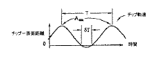
Tapping mode (Intermittent Contact) emerged as a solution to the sample damage problem by oscillating the cantilever near its resonant frequency, reducing lateral forces significantly. While this mode provides excellent resolution for many applications, it struggles with capturing rapid dynamic processes and can still exert excessive forces on extremely delicate samples.
Non-contact mode operates with the tip hovering above the sample surface, detecting long-range forces without physical contact. This approach minimizes sample damage but typically delivers lower resolution and is highly susceptible to environmental noise, limiting its practical applications in standard laboratory conditions.
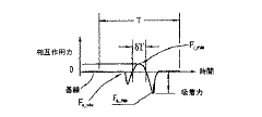
Peak Force Tapping represents a newer advancement that precisely controls tip-sample interaction forces by performing force curves at each pixel. This mode offers superior force control but requires sophisticated hardware and complex parameter optimization, creating a steep learning curve for new users.
Multifrequency AFM techniques have emerged to capture multiple sample properties simultaneously by exciting and monitoring multiple cantilever resonance modes. While promising, these methods generate complex datasets requiring advanced processing algorithms and specialized expertise for meaningful interpretation.
A significant technical challenge across all modes is the parameter selection problem. Operators must optimize numerous interdependent variables including scan rate, setpoint, gain settings, and tip characteristics based on sample properties that may be initially unknown. This creates a circular problem where optimal imaging requires knowledge of the very properties being investigated.
Environmental factors present additional challenges, as temperature fluctuations, acoustic vibrations, and electromagnetic interference can dramatically affect measurement quality. Advanced isolation systems address these issues but add considerable cost and complexity to AFM setups.
Tip degradation during scanning represents another persistent challenge, as tip geometry changes affect resolution and force application. Current solutions involving frequent tip replacement or characterization interrupt workflow and increase operational costs significantly.
Contact mode, the original AFM implementation, offers high-resolution topographical imaging but suffers from significant lateral forces that can damage delicate samples and distort measurements. This mode remains valuable for robust samples but presents considerable limitations for soft biological specimens and nanomaterials.
Tapping mode (Intermittent Contact) emerged as a solution to the sample damage problem by oscillating the cantilever near its resonant frequency, reducing lateral forces significantly. While this mode provides excellent resolution for many applications, it struggles with capturing rapid dynamic processes and can still exert excessive forces on extremely delicate samples.
Non-contact mode operates with the tip hovering above the sample surface, detecting long-range forces without physical contact. This approach minimizes sample damage but typically delivers lower resolution and is highly susceptible to environmental noise, limiting its practical applications in standard laboratory conditions.
Peak Force Tapping represents a newer advancement that precisely controls tip-sample interaction forces by performing force curves at each pixel. This mode offers superior force control but requires sophisticated hardware and complex parameter optimization, creating a steep learning curve for new users.
Multifrequency AFM techniques have emerged to capture multiple sample properties simultaneously by exciting and monitoring multiple cantilever resonance modes. While promising, these methods generate complex datasets requiring advanced processing algorithms and specialized expertise for meaningful interpretation.
A significant technical challenge across all modes is the parameter selection problem. Operators must optimize numerous interdependent variables including scan rate, setpoint, gain settings, and tip characteristics based on sample properties that may be initially unknown. This creates a circular problem where optimal imaging requires knowledge of the very properties being investigated.
Environmental factors present additional challenges, as temperature fluctuations, acoustic vibrations, and electromagnetic interference can dramatically affect measurement quality. Advanced isolation systems address these issues but add considerable cost and complexity to AFM setups.
Tip degradation during scanning represents another persistent challenge, as tip geometry changes affect resolution and force application. Current solutions involving frequent tip replacement or characterization interrupt workflow and increase operational costs significantly.
Parameter Selection Strategies for Different AFM Modes
01 Advanced AFM probe technologies
Innovations in atomic force microscopy probe design enable more precise imaging capabilities. These include specialized tip geometries, materials, and coatings that enhance resolution and reduce artifacts. Advanced probes can better interact with sample surfaces at the nanoscale, providing clearer topographical data and improved measurement accuracy while minimizing sample damage during scanning.- Advanced AFM probe technologies: Specialized probe designs enhance atomic force microscopy imaging precision. These include probes with optimized tip geometries, novel materials, and modified cantilevers that improve resolution and reduce artifacts. Advanced probes enable more accurate surface topography measurements and can be tailored for specific sample types, allowing for nanometer-scale precision in various research and industrial applications.
- AFM operational modes and techniques: Various operational modes and techniques have been developed to enhance atomic force microscopy imaging precision. These include tapping mode, non-contact mode, and force mapping approaches that minimize sample damage while maximizing resolution. Advanced techniques incorporate feedback control systems and signal processing algorithms to improve image quality, reduce noise, and enable measurement of additional sample properties beyond topography.
- Environmental control systems for AFM: Environmental control systems significantly improve atomic force microscopy imaging precision by minimizing external disturbances. These systems regulate temperature, humidity, and vibration, creating stable conditions for high-resolution imaging. Vacuum chambers, acoustic isolation, and temperature-controlled stages help eliminate artifacts and drift, enabling more consistent and reproducible measurements at the atomic scale.
- Data processing and image enhancement algorithms: Sophisticated data processing and image enhancement algorithms are crucial for achieving precise atomic force microscopy imaging. These computational methods include noise filtering, drift correction, and artifact removal techniques that improve image quality. Advanced algorithms can reconstruct surface features with higher fidelity, apply statistical analysis to raw data, and enhance contrast to reveal subtle nanoscale structures that might otherwise be obscured.
- Hybrid and multimodal AFM systems: Hybrid and multimodal atomic force microscopy systems combine AFM with complementary techniques to enhance imaging precision. These integrated approaches may incorporate optical microscopy, Raman spectroscopy, or electrical measurements to provide correlated multidimensional data. By simultaneously capturing different physical properties, these systems offer more comprehensive sample characterization and improved contextual understanding of nanoscale features.
02 AFM operational mode enhancements
Various operational modes have been developed to optimize atomic force microscopy imaging for different applications. These include tapping mode, non-contact mode, and specialized scanning techniques that reduce sample damage while increasing resolution. These operational enhancements allow for better control of the probe-sample interaction forces, resulting in more precise imaging of delicate biological samples, semiconductor materials, and other nanoscale structures.Expand Specific Solutions03 Signal processing and noise reduction techniques
Advanced signal processing algorithms and noise reduction techniques significantly improve the quality of atomic force microscopy images. These methods filter out environmental vibrations, thermal drift, and electronic noise that can degrade image quality. Real-time data processing, adaptive filtering, and statistical analysis techniques enhance contrast and resolution, allowing for more accurate representation of surface features at the atomic scale.Expand Specific Solutions04 Environmental control systems for AFM
Environmental control systems for atomic force microscopy enable precise imaging under controlled conditions. These systems regulate temperature, humidity, and atmospheric composition during scanning, reducing thermal drift and minimizing contamination. Vacuum, liquid, and gas-controlled chambers allow for specialized imaging environments that preserve sample integrity and enhance measurement stability, resulting in higher resolution images and more reproducible results.Expand Specific Solutions05 Integration with complementary imaging techniques
Combining atomic force microscopy with other imaging and analytical techniques creates powerful hybrid systems for comprehensive sample characterization. These integrated approaches merge AFM with optical microscopy, Raman spectroscopy, or electron microscopy to correlate topographical data with chemical, electrical, or mechanical properties. Such multimodal imaging provides deeper insights into sample characteristics while maintaining the high spatial resolution inherent to AFM.Expand Specific Solutions
Leading AFM Manufacturers and Research Groups
Atomic Force Microscopy (AFM) technology is currently in a mature growth phase, with an expanding market driven by increasing applications in materials science, nanotechnology, and life sciences. The global AFM market is estimated to be valued at approximately $500-600 million, with steady annual growth of 5-7%. The competitive landscape features established industry leaders like Bruker Nano, Oxford Instruments Asylum Research, and Nanosurf AG, who dominate with comprehensive product portfolios and advanced imaging capabilities. Academic institutions including Beihang University, Zhejiang University, and University of Maryland are driving innovation through research partnerships. The technology demonstrates high maturity with ongoing refinements in imaging parameters, resolution capabilities, and specialized modes, while companies like FEI Co. and Veeco Instruments are expanding applications through integration with complementary technologies and development of parameter optimization solutions.
Bruker Nano, Inc.
Technical Solution: Bruker Nano has developed PeakForce Tapping technology for atomic force microscopy that enables simultaneous acquisition of multiple sample properties while protecting both tip and sample. Their system automatically optimizes imaging parameters through real-time force curve analysis at each pixel, maintaining constant peak force interaction. The technology incorporates ScanAsyst® which automatically adjusts key parameters including setpoint, feedback gains, and scan rate based on image quality metrics. For precise imaging, Bruker's systems offer multiple specialized modes including PeakForce QNM® (Quantitative Nanomechanical Mapping) that provides mechanical property mapping with nanoscale resolution, PeakForce TUNA™ for electrical characterization, and PeakForce KPFM™ for surface potential measurements. Their FastScan technology enables high-speed imaging while maintaining resolution through optimized scanner design and advanced feedback algorithms[1][2].
Strengths: Automated parameter optimization reduces user expertise requirements and improves reproducibility. Multiple specialized modes allow comprehensive sample characterization beyond topography. High-speed imaging capabilities maintain resolution while increasing throughput. Weaknesses: Proprietary technology creates vendor lock-in. Higher cost compared to basic AFM systems. Some specialized modes require specific probe types, increasing operational costs.
Veeco Instruments, Inc.
Technical Solution: Veeco Instruments has pioneered the BlueDrive™ photothermal excitation technology for atomic force microscopy, which provides pure cantilever excitation by directly driving the cantilever photothermally rather than through acoustic excitation. This approach eliminates spurious resonances and fluid coupling effects, resulting in cleaner frequency spectra and more accurate measurements. Their Dimension FastScan system incorporates adaptive scanning technology that automatically adjusts scan parameters based on sample topography and feedback signals. For precise imaging parameter selection, Veeco has developed PeakForce QNM® (Quantitative Nanomechanical Mapping) mode that allows simultaneous acquisition of nanomechanical properties including modulus, adhesion, and deformation while maintaining low forces to protect delicate samples. The system features closed-loop scanners with position sensors that provide positional accuracy and eliminate hysteresis and creep effects common in piezoelectric scanners[3][4].
Strengths: Photothermal excitation provides cleaner frequency response, especially critical for liquid imaging. Closed-loop scanning offers superior positional accuracy and repeatability. Adaptive scanning technology reduces operator intervention requirements. Weaknesses: Higher system complexity increases maintenance requirements. Premium pricing positions technology beyond reach of some research budgets. Some advanced modes require specialized probes that increase operational costs.
Critical Parameters Affecting AFM Image Resolution
Scanning probe microscope and method of operating the same
PatentActiveJP2018036272A
Innovation
- The implementation of Peak Force Tapping (PFT) Mode, which uses instantaneous interaction force control to minimize parasitic coupling and automate feedback adjustments, allowing for high-resolution imaging with minimal user intervention and reduced tip-sample forces.
Atomic force microscopy controller and method
PatentWO2012115653A1
Innovation
- A controller system that determines and adjusts its output based on the loop response for both on-surface and off-surface movements of the cantilever, using notch filters to mitigate off-surface resonance conditions and maintain stability, thereby preventing instability and improving image resolution.


Sample Preparation Protocols for Optimal AFM Imaging
Sample preparation is a critical determinant of successful atomic force microscopy (AFM) imaging outcomes. The quality of sample preparation directly impacts the resolution, accuracy, and reproducibility of AFM measurements. For biological samples, fixation techniques must preserve structural integrity while minimizing artifacts. Chemical fixation using glutaraldehyde or formaldehyde stabilizes proteins, while physical methods like rapid freezing are preferred for preserving dynamic cellular structures.
Surface cleanliness represents a fundamental requirement across all sample types. Contaminants at the nanoscale can significantly distort imaging results, necessitating rigorous cleaning protocols. For hard materials, sequential ultrasonic cleaning in solvents of decreasing polarity effectively removes organic and inorganic contaminants. Plasma cleaning offers an additional step for removing persistent organic residues without physical damage to the sample surface.
Substrate selection must be optimized based on the specific AFM mode and sample characteristics. Atomically flat substrates like mica, highly oriented pyrolytic graphite (HOPG), or silicon wafers provide ideal backgrounds for high-resolution imaging. For biological specimens, functionalized substrates that promote adhesion without denaturing biomolecules are essential. Poly-L-lysine coated surfaces enhance electrostatic interactions with negatively charged biological samples.
Immobilization strategies vary by sample type and imaging environment. For liquid-phase imaging of biological samples, gentle chemical crosslinking or physisorption techniques maintain native conformations. Polymer samples often require spin-coating or solution casting to achieve uniform thickness. Nanoparticles and other discrete structures benefit from electrostatic immobilization or chemical tethering to prevent displacement during scanning.
Environmental control during preparation significantly impacts sample stability. Temperature, humidity, and atmospheric composition must be monitored and controlled to prevent sample degradation. For hygroscopic materials, preparation under controlled humidity prevents morphological changes. Oxygen-sensitive samples require preparation under inert atmospheres to prevent oxidative damage.
Final preparation steps should include careful consideration of imaging conditions. For contact mode AFM, samples must withstand lateral forces without deformation. Tapping mode requires consideration of resonance effects on delicate structures. Force mapping applications demand consistent mechanical properties across the sample surface. Calibration standards prepared alongside experimental samples provide crucial reference points for quantitative measurements and instrument performance verification.
Data Processing and Image Analysis Methods
Data processing and image analysis are critical components in atomic force microscopy (AFM) that significantly impact the quality and interpretability of results. Raw AFM data typically contains various artifacts and noise that must be addressed through sophisticated processing algorithms. The primary processing steps include flattening to remove sample tilt and bow distortions, which is accomplished through polynomial fitting of scan lines or planes. This correction is essential for accurate topographical representation, particularly when imaging samples with nanoscale features.
Filtering techniques play a crucial role in enhancing image quality by reducing noise while preserving important structural information. Commonly employed methods include Gaussian filters for general noise reduction, median filters for eliminating outlier data points, and Fourier transform-based filters for addressing periodic noise patterns. The selection of appropriate filtering parameters requires careful consideration to avoid introducing artificial features or removing genuine sample characteristics.
Advanced image analysis methods have evolved significantly with the integration of machine learning algorithms. These computational approaches enable automated feature recognition, statistical analysis of surface properties, and classification of different material phases within heterogeneous samples. Particularly valuable in complex biological and composite material studies, these techniques allow for quantitative characterization of surface roughness, particle size distribution, and domain structures.
Three-dimensional reconstruction from AFM data represents another critical analytical capability. By processing height information across the scanning area, researchers can generate volumetric representations that provide insights into sample morphology not immediately apparent in traditional 2D presentations. This approach is particularly valuable when examining biological structures, polymer assemblies, and nanofabricated devices where spatial relationships are complex.
Correlation analysis between different AFM channels (height, phase, amplitude) offers multidimensional insights into sample properties. By examining relationships between mechanical, electrical, and topographical data, researchers can identify material boundaries, detect subsurface features, and characterize compositional variations. This multimodal analysis is especially powerful when combined with statistical methods that can identify patterns and anomalies across large datasets.
Batch processing capabilities have become increasingly important as AFM applications expand to high-throughput screening and quality control environments. Automated workflows that can process multiple images with consistent parameters enable efficient analysis of large sample sets, facilitating comparative studies and statistical validation of observed phenomena. These capabilities are essential for industrial applications where reproducibility and throughput are paramount considerations.
Unlock deeper insights with Patsnap Eureka Quick Research — get a full tech report to explore trends and direct your research. Try now!
Generate Your Research Report Instantly with AI Agent
Supercharge your innovation with Patsnap Eureka AI Agent Platform!