
Microscope image cell classification judgment method
A technology of cell classification and microscopy, applied in image analysis, capturing objects visible under the microscope, image enhancement, etc., can solve the problems affecting the classification and judgment of pathological somatic cell cancers, the lack of identification and diagnosis of cell-related lesions, and the impact on sampling of cancer patients Detect and observe the efficiency and other issues to achieve the effect of satisfying the ability of traceability, improving the ability of cell classification and judgment, and reducing the difficulty of classification
Patent Information
- Authority / Receiving Office
- CN · China
- Patent Type
- Applications(China)
- Current Assignee / Owner
- Publication Date
- 2021-07-30
Smart Images
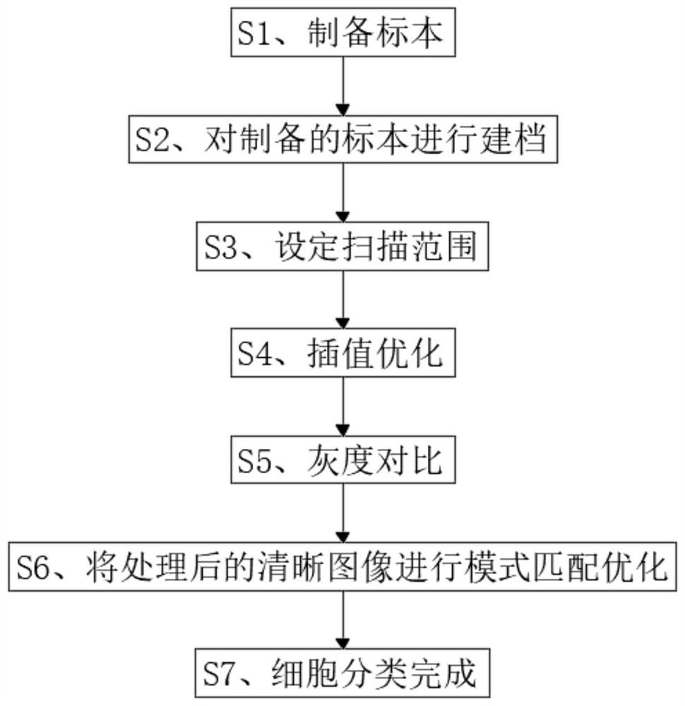
Figure 1
Abstract
Description
technical field
[0001] The invention belongs to the technical field of cell classification and judgment, and in particular relates to a judgment method for cell classification of microscope images. Background technique
[0002] Today, with the development of medical technology, early cancer prevention is the main prevention method for cancer. In order to determine whether cystic polyps are cancer or precursors, it is generally necessary to sample cystic polyps for observation to ensure whether they are diffuse cancer cells.
[0003] When sampling and observing cells, it is generally necessary for inspectors to compare and subdivide cells based on their own clinical experience. The efficiency of judging cell classification under a microscope is low, which affects the ability to quickly screen cancer cells and cannot be linked with the medical system. To cooperate, physicians need to add cell judgment and classification results in the system by themselves. The lack of identifi...
Examples
Embodiment 1
[0039] The invention provides a technical solution: take a clean glass slide, absorb physiological saline and add it dropwise to the center of the glass slide, take cells detached from the body and place them in the normal saline, dry the slide or heat it with alcohol light to dry the slide, absorb Put the fixative into the glass slide and fix it for 10 minutes, absorb the corresponding Papanicolaou staining agent by Papanicolaou staining method for 2 minutes, rinse off the excess dye, and keep it for later use. The image cell classification observation hardware system calculates the area of the specimen through the hardware system, sets the scanning range according to the specimen size, controls the scanning focus mode, obtains the cell scanning image after scanning the specimen, and double-checks the scanned cell image. Cubic interpolation optimization, algorithmic processing of the cell image in the scanned image to obtain a smoother image edge, and at the same time enlarg...
Embodiment 2
[0050] Take a clean glass slide, absorb physiological saline and add it dropwise to the center of the glass slide, take the detached cells from the body and put them in the normal saline, dry them in the air or heat them with alcohol lamps to dry the slides, absorb the fixative and place them on the slide Fix for 10 minutes, absorb the corresponding Papanicolaou staining agent for 2 minutes by Papanicolaou staining method, rinse off the excess dye, and keep it for later use. After the hardware system calculates the area of the specimen, the scanning range is set according to the size of the specimen, and the scanning focus mode is controlled. Algorithm processing is performed on the cell image to obtain a smoother image edge, and at the same time, the proportion of the cell image in the image is enlarged, invalid black borders and blank areas are removed, and the contrast pixels of the enlarged image are optimized and retained. After the clearest image in the first scanned c...
Embodiment 3
[0054] The difference from both Examples 1 and 2 is that, take a clean glass slide, absorb physiological saline and add it dropwise to the center of the slide, take the detached cells from the body and place them in normal saline, dry them in the air or heat them with an alcohol lamp to make the slides After drying, absorb the fixative solution and put it on the glass slide, fix it for 10 minutes, absorb the corresponding Papanicolaou staining agent for 2 minutes, rinse off the excess dye, and keep it for later use. Connect the image cell classification and observation hardware system under the microscope. After calculating the area of the specimen through the hardware system, set the scanning range according to the size of the specimen, control the scanning focus mode, and obtain the cell scanning image after scanning the specimen. The image is optimized by bicubic interpolation, and the cell image in the scanned image is processed by an algorithm to obtain a smoother image ...