
Tissue grasping devices and related methods
a technology of tissue and stent, applied in the field of tissue grasping devices, can solve the problems of more difficult to grasp symmetrically in the same grasp as the other leaflet, and achieve the effects of optimal coapted configuration, optimal coapted configuration, and optimal coapted configuration
Patent Information
- Authority / Receiving Office
- US · United States
- Patent Type
- Applications(United States)
- Current Assignee / Owner
- Publication Date
- 2019-05-16
Smart Images
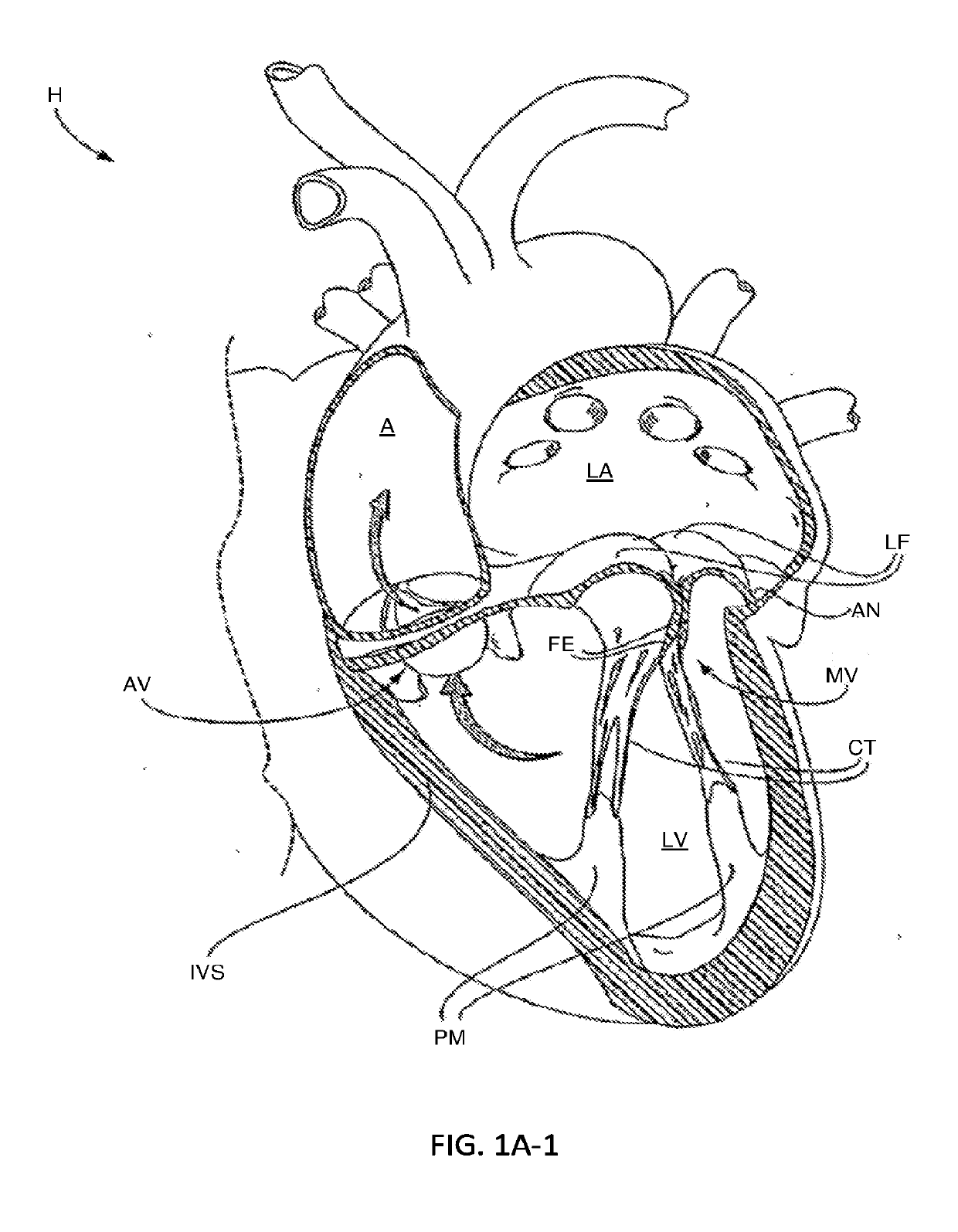
Figure 1 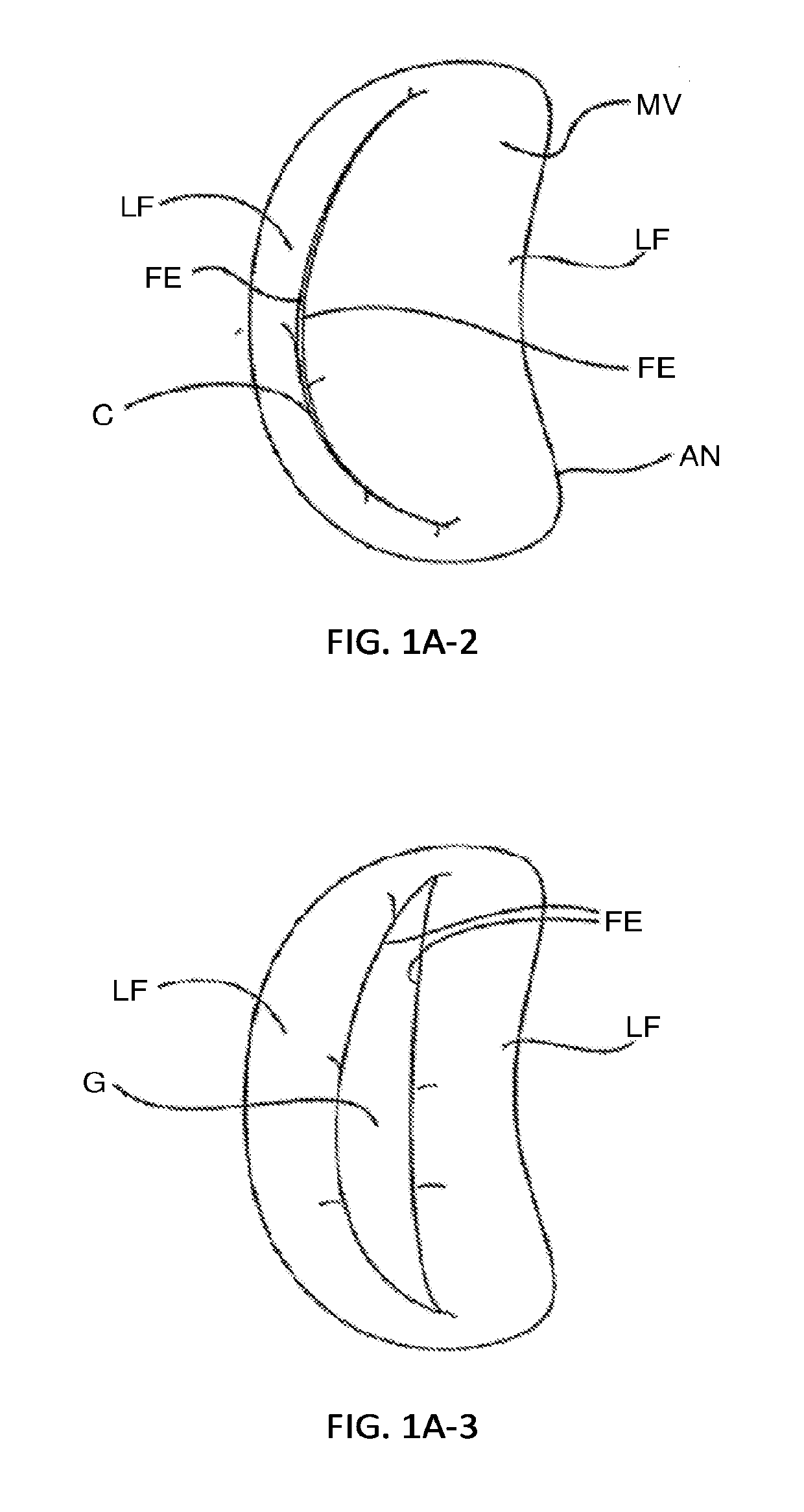
Figure 2 
Figure 3
Abstract
Description
CROSS-REFERENCE TO RELATED APPLICATIONS
[0001] This application is a continuation of PCT Application No. PCT / US17 / 42003 (Attorney Docket No. 52206-703.601), filed Jul. 13, 2017, which claims the benefit of Provisional Application No. 62 / 361,953 (Attorney Docket No. 52206-703.101), filed Jul. 13, 2016, the entire content of which is incorporated herein by reference; this application also claims the benefit of Provisional Application No. 62 / 617,946 (Attorney Docket No. 52206-704.101), filed Jan. 16, 2018, the entire content of which is incorporated herein by reference.BACKGROUND OF THE INVENTION1. Field of the Invention
[0002] The present invention relates generally to medical methods, devices, and systems. In particular, the present invention relates to methods, devices, and systems for the endovascular, percutaneous, or minimally invasive surgical treatment of bodily tissues, such as tissue approximation or valve repair. More particularly, the present invention relates to methods and de...
Examples
Embodiment Construction
I. Cardiac Physiology
[0153]The left ventricle LV of a normal heart H in systole is illustrated in FIG. 1A-1. The left ventricle LV is contracting and blood flows outwardly through the tricuspid (aortic) valve AV in the direction of the arrows. Back flow of blood or “regurgitation” through the mitral valve MV is prevented since the mitral valve is configured as a “check valve” which prevents back flow when pressure in the left ventricle is higher than that in the left atrium LA. The mitral valve MV comprises a pair of leaflets having free edges FE which meet evenly to close, as illustrated in FIG. 1A-1. The opposite ends of the leaflets LF are attached to the surrounding heart structure along an annular region referred to as the annulus AN. The free edges FE of the leaflets LF are secured to the lower portions of the left ventricle LV through chordae tendineae CT (referred to hereinafter as the chordae) which include plurality of branching tendons secured over the lower surfaces of e...