
Computed tomography imaging
a computed tomography and imaging technology, applied in the field of computed tomography imaging, can solve the problems of low resolution of conventional two-dimensional x-ray imaging, and achieve the effects of high resolution, low dose, and high resolution
Patent Information
- Authority / Receiving Office
- US · United States
- Patent Type
- Applications(United States)
- Current Assignee / Owner
- Publication Date
- 2022-01-20
Smart Images
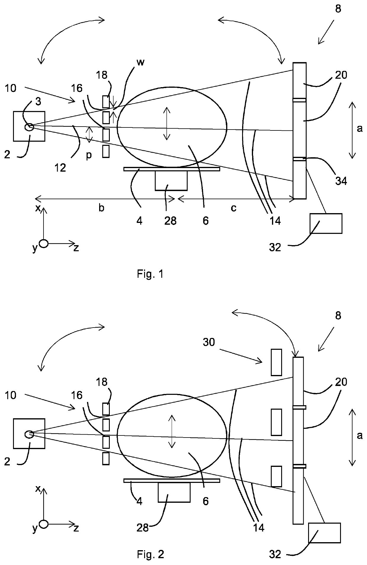
Figure 1 
Figure 2 
Figure 3
Abstract
Description
[0001] The invention relates to a method of computed tomography (CT) imaging and apparatus for carrying out CT imaging.BACKGROUND
[0002] X-Ray CT has become a very important technique for diagnostic imaging, especially in the field of medicine. However, the technique does involve exposing the subject to ionising X-ray radiation. The technique generally results in lower resolution than conventional two dimensional X-ray imaging, such as mammography or standard X-ray imaging. The reason for the lower resolutions achieved is that to increase an image resolution in two dimensions there is normally a quadratic increase in dose—so to double the resolution the dose must be multiplied by 4. An increase in resolution in three dimensions leads to a cubic increase in dose. Therefore, to keep doses to reasonable levels lower resolutions are used than for conventional two dimensional X-ray imaging which has significant implications for what CT can and cannot detect and resolve. This in turn can hav...
Examples
Embodiment Construction
[0072]The CT apparatus comprises an X-ray source 2 having a focal spot 3, a subject stage 4 for supporting a subject 6 such as a human being or a tissue sample, and an X-ray detector 8 in the form of a two dimensional pixel detector having a plurality of pixels 20 of pixel size a. The subject stage is not fixed in position as will be described in more detail below. Individual pixels 20 are separated by regions 34.
[0073]A mask 10 is provided having a plurality of apertures 16 of width w at a mask period p, the apertures being between block regions 18 in the form of septa. The beam 12 emitted by the X-ray source 2 is broken up into a plurality of beamlets 14 by the mask 10, each beamlet being generated by a respective aperture 16. The mask period p matches the detector pixel size a in that p=a / m where m is the magnification between mask and detector. In other words, each pixel 20 receives a respective beamlet 14.
[0074]A processing apparatus 32 is connected to the X-ray detector 8 for ...