
Device and method for optical imaging of retinal function
a retinal function and optical imaging technology, applied in the field of retinal neuronal activity imaging, can solve the problems of difficult monitoring changes, perimetry is relatively insensitive to early detection, optic nerve damage, etc., and achieve the effect of increasing the signal-to-noise ratio (snr) and increasing the sensitivity of the instrumen
Patent Information
- Authority / Receiving Office
- US · United States
- Patent Type
- Patents(United States)
- Current Assignee / Owner
- Publication Date
- 2006-10-10
- Estimated Expiration
- Not applicable · inactive patent
Smart Images
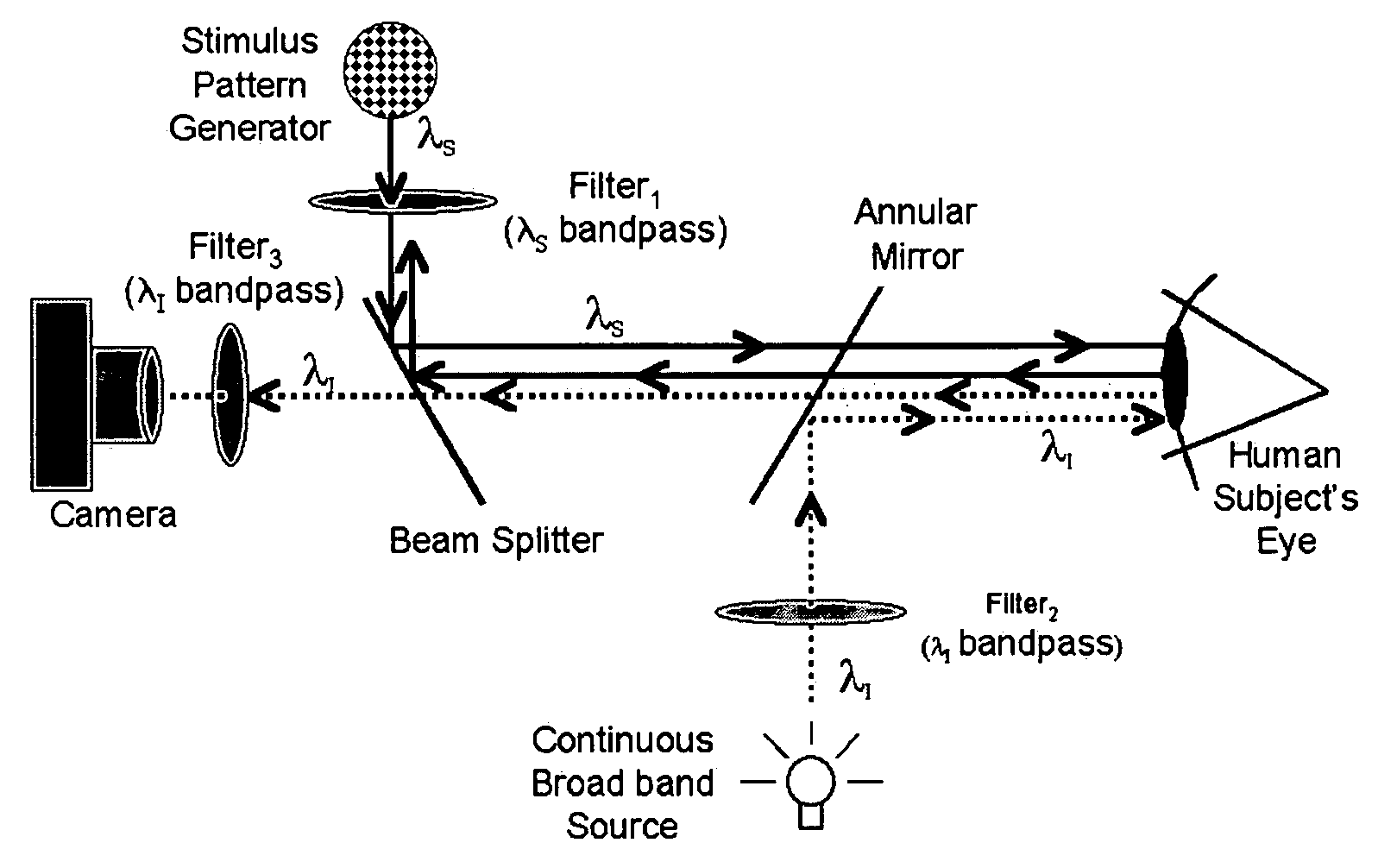
Figure 1 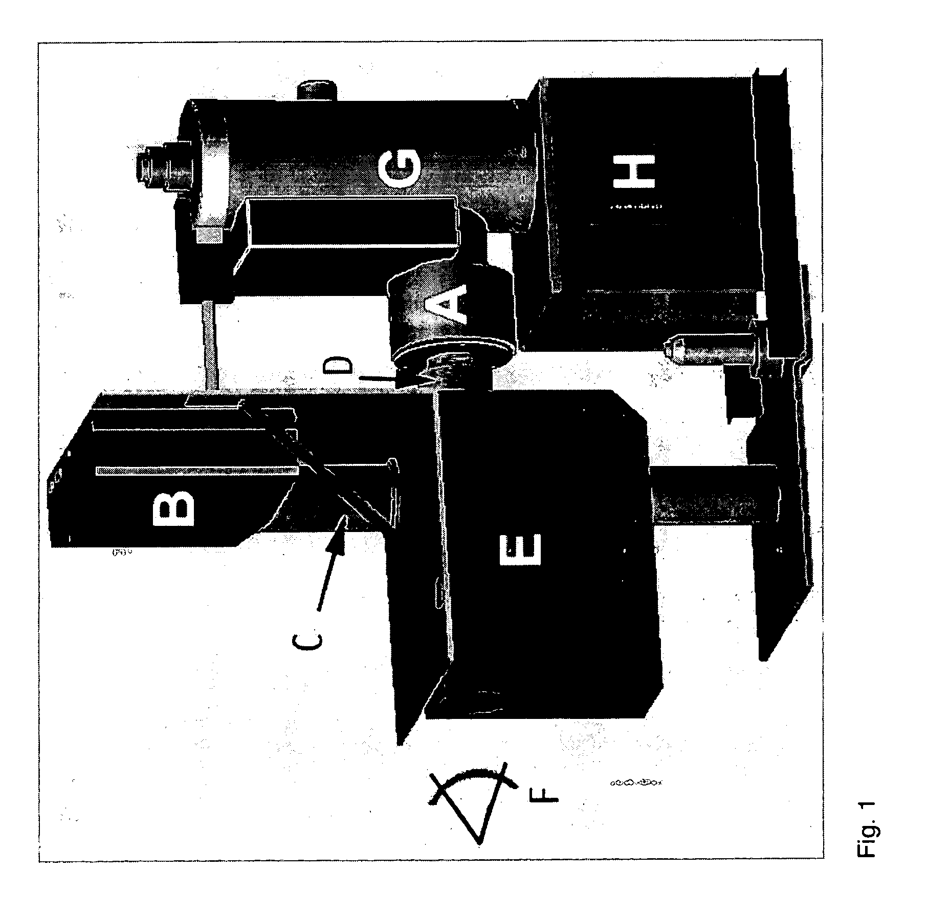
Figure 2 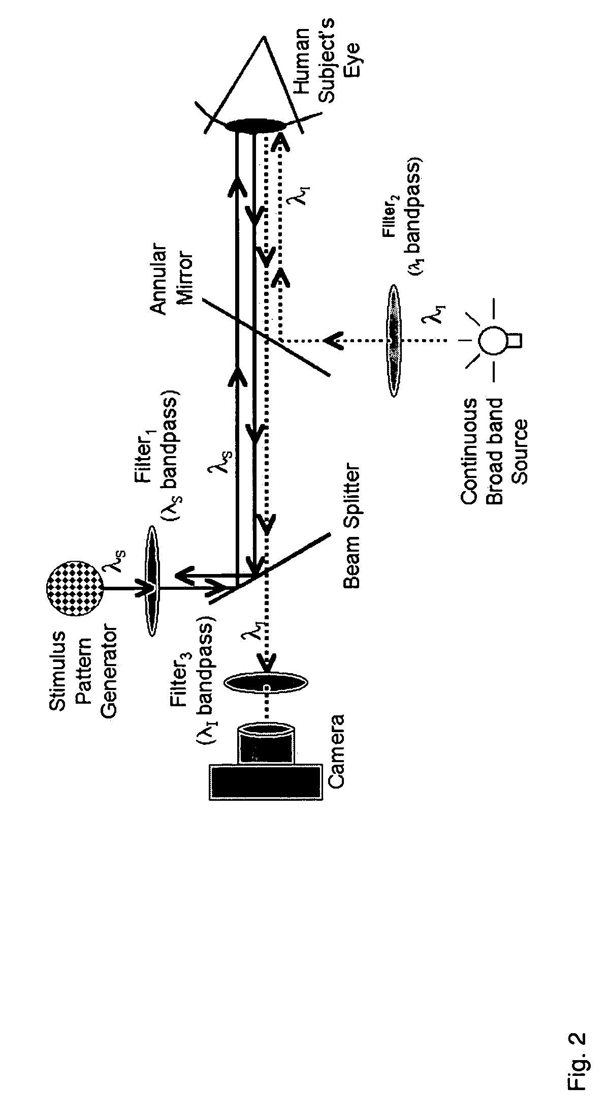
Figure 3
Abstract
Description
CROSS-REFERENCE TO RELATED APPLICATION
[0001] The benefit of the filing date of U.S. Provisional Patent Application Ser. No. 60 / 349,435, filed Jan. 18, 2002, entitled “OPTICAL IMAGING DEVICE OF RETINA FUNCTION,” is hereby claimed, and the specification thereof incorporated herein in its entirety.ACKNOWLEDGMENT
[0002] This invention was made with government support under Grant number 1R43EY12915-01A1 awarded by the National Eye Institute. The government has certain rights in the invention.FIELD OF THE INVENTION
[0003] The invention relates in general to retinal neuronal activity imaging. More particularly, the invention relates to instrumentation and methodologies in the diagnostic imaging of stimulus responsive ocular fundus neuronal activity.BACKGROUND OF THE INVENTION
[0004] The current standard for detecting and monitoring progression of diseases of the optic nerve (i.e. glaucoma, ischemic optic neuropathy, compressive optic neuropathy) and retina is visual field testing (perimetry). Per...
Examples
Embodiment Construction
[0037]The optical recording of intrinsic neuronal signals is suited for objective assessment of retinal function. Like the cortex, neuronal signals from the retina can be recorded from a large area at once with high spatial resolution. Unlike cortex imaging, retinal imaging can be acquired directly through a dilated pupil or other area of the eye using an appropriate fundus imaging system. Optical recording of retinal function is noninvasive and ideal for clinical application.
[0038]Optical functional recording is possible in the retina because the retina is a direct extension of the brain and part of the central nervous system. Neuronal activity of the retina is fundamentally similar to that of the brain. Like the brain, appropriate metabolic changes (changes in hemoglobin oxygen saturation and state of tissue cytochrome for example) can be detected in the retina in response to changes in inspired oxygen levels.
[0039]The following steps are included in a preferred embodiment of an i...