
Agglutination assay
a technology of agglutination and assay, which is applied in the field of agglutination assay, can solve the problems of difficult to achieve stable agglutination with smaller analytes
Patent Information
- Authority / Receiving Office
- US · United States
- Patent Type
- Patents(United States)
- Current Assignee / Owner
- Publication Date
- 2015-12-01
- Estimated Expiration
- Not applicable · inactive patent
Smart Images
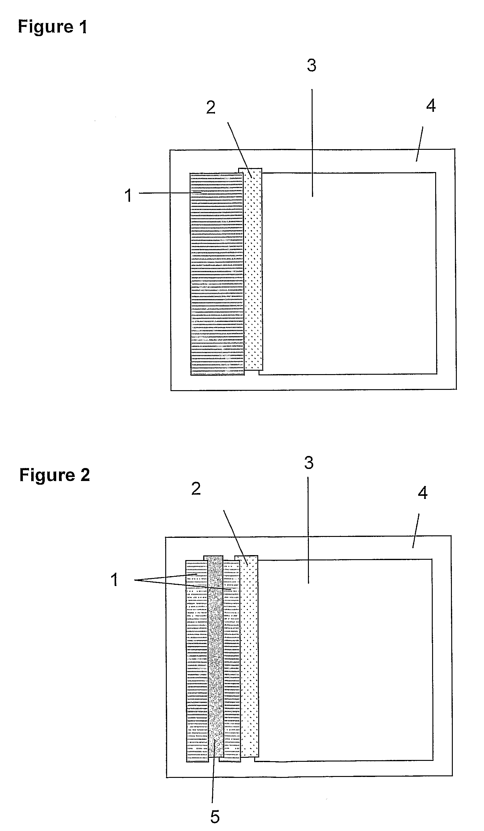
Figure 1 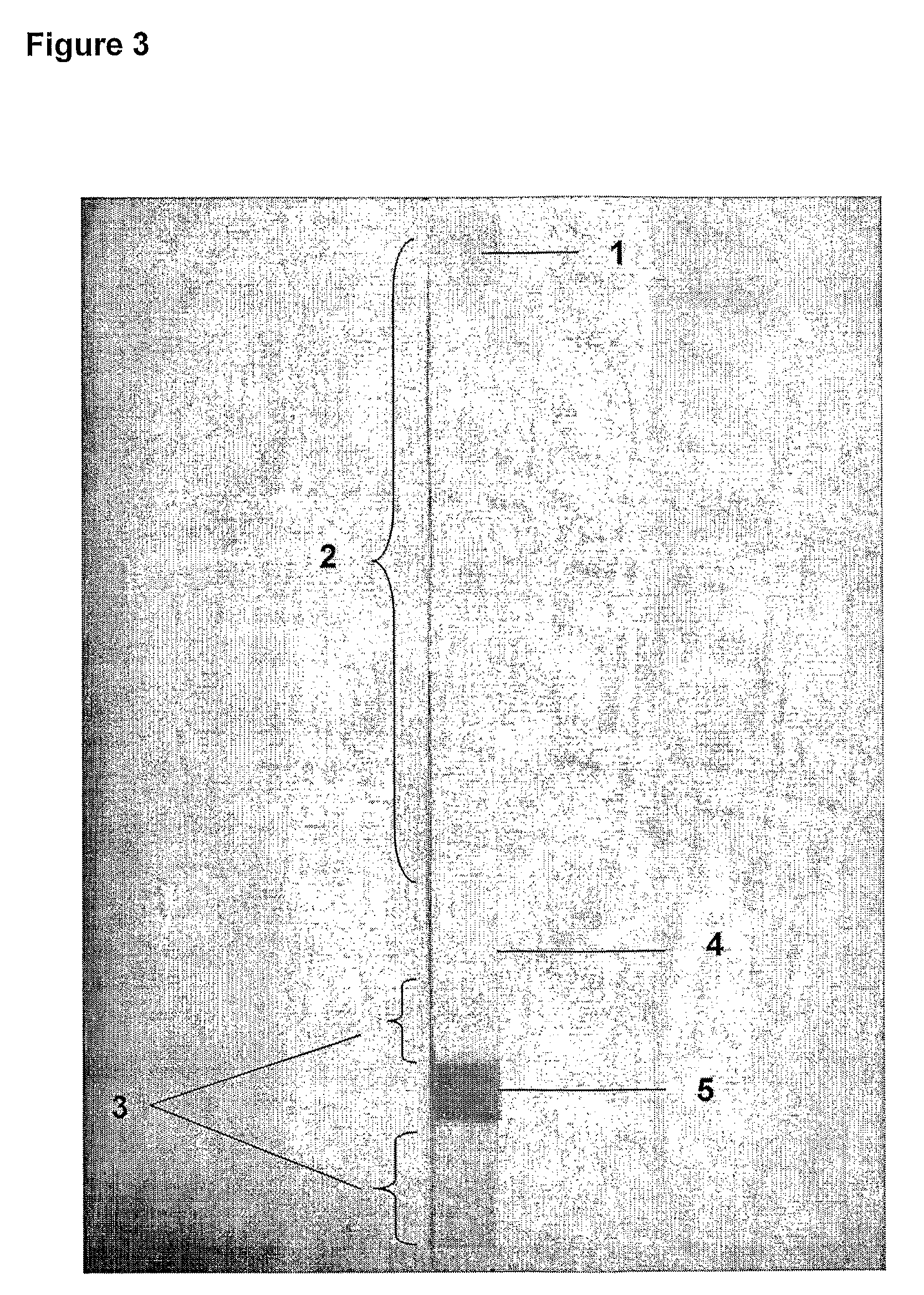
Figure 2 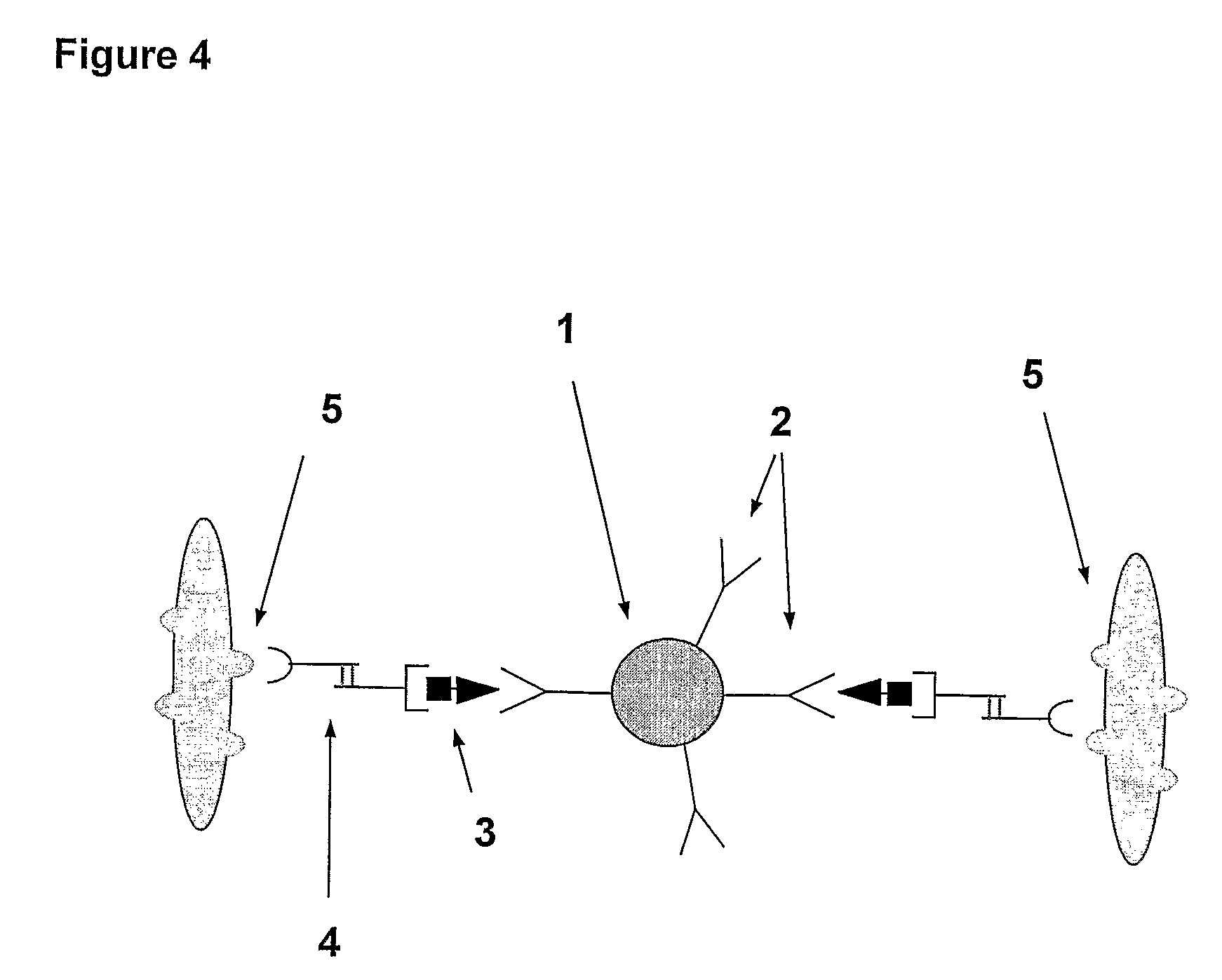
Figure 3
Abstract
Description
REFERENCE TO RELATED APPLICATIONS
[0001] This application is the US national phase entry of International Patent Application No. PCT / GB2006 / 004204, filed Nov. 13, 2006, which claims priority to UK Patent Applications No. 0523124.6, filed Nov. 12, 2005 and 0610973.0 filed Jun. 3, 2006.FIELD OF THE INVENTION
[0002] The present invention relates to an agglutination assay for detection of analytes in a sample. In particular, the invention relates to porous carrier-based assays and assay devices, kits comprising means for conducting such assays, and assay methods for the detection of analyte in a sample.BACKGROUND TO THE INVENTION
[0003] Immunoassays are a well established technique for detecting and quantifying analytes in samples. They are particularly useful for detecting and / or measuring substances in biological samples as an aid to disease diagnosis and prognosis, and for predicting a patient's response to therapy. Techniques such as the radioimmunoassay and enzyme immunoassay, which revo...
Examples
example 1
Preparation of Soluble Hub Reagent 1
[0133]1. Desalt the anti-hCG (alpha-subunit) into 0.1 M phosphate pH 7.5 buffer, using a 1.6×15 cm G25M Sephadex column, and determine concentration and yield.
[0134]2. Activate the anti-hCG antibody, using 8 molar equivalents of NHS-PEG-MAL. Incubate the reaction mixture at 20° C. for two hours. Quench the reaction with 100 molar equivalents of glycine and desalt the maleimide-activated anti-hCG into 5 mM EDTA, PBS pH 7.3 buffer using two shots down a 1.6×15 cm G50F Sephadex column. Determine concentration and yield of activated antibody.
[0135]3. Activate a 500 kDalton aminodextran using 1000 molar equivalents of 2-Iminothiolane (2-IT). Incubate the reaction mixture at 20° C. for 110 minutes. Desalt the thiol activated aminodextran into 5 mM EDTA, PBS pH 7.3 buffer, using G25M Sephadex media. Determine incorporation ratio of thiol:aminodextran using the Ellman's assay.
[0136]4. Add 25 Molar equivalents of the maleimide-activated anti-hCG antibody t...
example 2
Preparation of Soluble Hub Reagent 2
[0137]1. Desalt the anti-hCG (alpha-subunit) into 0.1 M phosphate pH 7.5 buffer, using a 1.6×15 cm G25M Sephadex column, and determine concentration and yield.
[0138]2. Activate the anti-hCG antibody, using 8 molar equivalents of NHS-PEG-MAL. Incubate the reaction mixture at 20° C. for two hours. Quench the reaction with 100 molar equivalents of glycine and desalt the maleimide-activated anti-hCG into 5 mM EDTA, PBS pH 7.3 buffer using two shots down a 1.6×15 cm G50F Sephadex column. Determine concentration and yield of activated antibody.
[0139]3. Activate a 500 kDalton aminodextran using 1000 molar equivalents of 2-Iminothiolane (2-IT). Incubate the reaction mixture at 20° C. for 110 minutes. Desalt the thiol activated aminodextran into 5 mM EDTA, PBS pH 7.3 buffer, using G25M Sephadex media. Determine incorporation ratio of thiol:aminodextran using the Ellman's assay.
[0140]4. Add 25 Molar equivalents of the maleimide-activated anti-hCG antibody t...
example 3
Preparation of Membrane Strips for Tests Using Wet Reagents
[0142]Membrane materials were cut to size as follows:[0143](i) Wick, e.g. Surewick G028-14 (Millipore), 30 mm×60 mm.[0144](ii) Agglutinate trapping membrane, e.g. Fusion 5 (Whatman), 5 mm×60 mm.[0145](iii) Absorbent sink, e.g. Absorbent Pad 222 (Ahlstrom), 55 mm×60 mm.[0146](iv) Self-adhesive plastic (×2) e.g. 0.04″ Clear polyester with D / C hydrophilic PSA (G&L) 70 mm×100 mm
[0147]A composite ‘card’ of the above materials was assembled as shown in FIG. 1. Adjacent membrane materials were overlapped by approximately 1 mm, to ensure good fluid transfer between successive sections of the strip. The second sheet of self-adhesive plastic was applied firmly to the upper surface. The resulting ‘card’ was sliced into 5 mm strips and the plastic trimmed to allow reagents and sample to enter the wick.