Method of reducing collateral flow in a portion of a lung

US20050288684A1Inactive Publication Date: 2005-12-29PNEUMRX
99 Cites 187 Cited by

Patent Information

Authority / Receiving Office
US · United States
Current Assignee / Owner
Publication Date
2005-12-29
Estimated Expiration
Not applicable · inactive patent

Smart Images

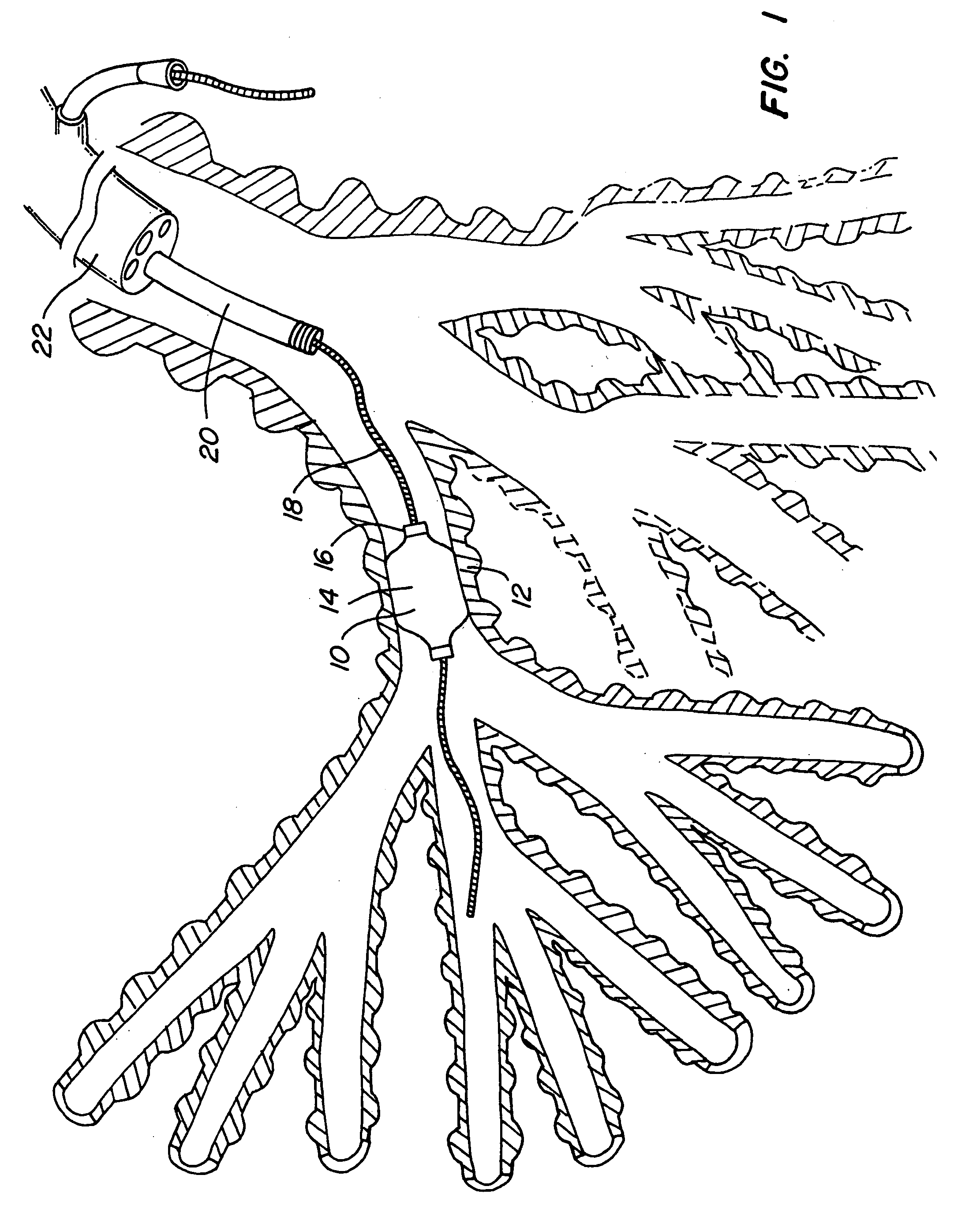
  • Figure 1
    Figure 1
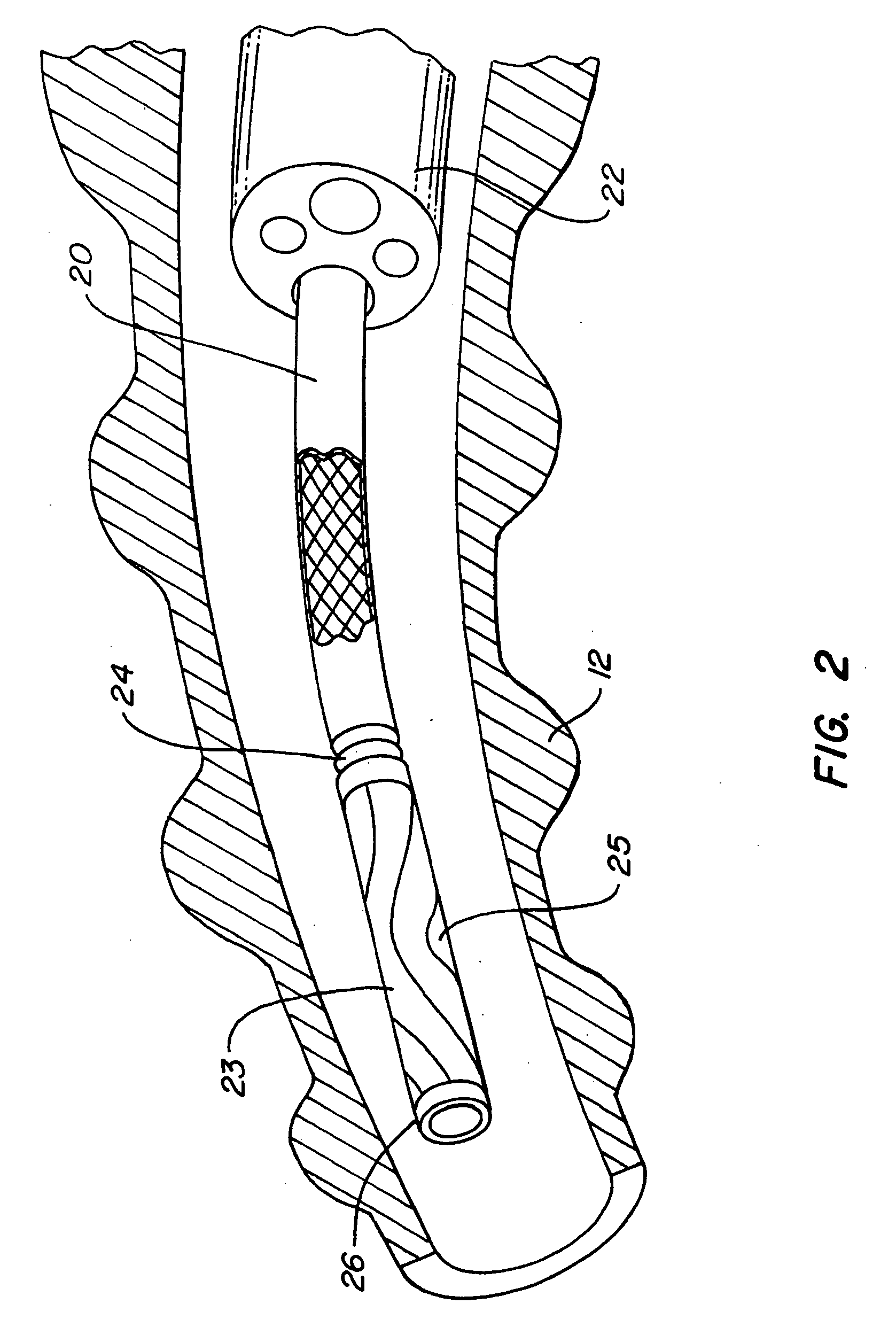
  • Figure 2
    Figure 2
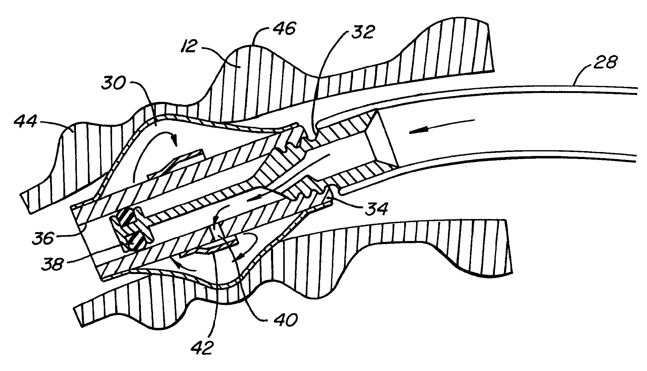
  • Figure 3
    Figure 3
Patent Text Reader

Abstract

This invention provides methods of treating a patient's lung. One aspect of the invention provides a method of treating a lung including the following steps: inserting a collateral flow blocking agent delivery device into an air passageway of the lung; and delivering a collateral flow blocking agent to reduce airflow between a portion of the lung distal to the delivery device and the exterior of the patient. Another aspect of the invention provides a method of treating a lung including the step of delivering a collateral flow blocking agent to reduce airflow through openings in lung tissue caused by tissue degradation.
Need to check novelty before this filing date? Find Prior Art

Description

CROSS-REFERENCE

[0001] This application claims the benefit of U.S. Provisional Application No. 60 / 580,565, filed Jun. 16, 2004, which is incorporated herein by reference in its entirety.BACKGROUND OF THE INVENTION

[0002] The primary role of the lung is to perform the function of breathing which assists in the intake of oxygen and removal of carbon dioxide from the body. The oxygen in air is inhaled through the mouth and trachea to the main bronchi. The bronchi divide at the end of the trachea into the left and right main bronchi and these respectively divide into bronchial branches, which “feed” the three lobes of the lung on the right and two on the left. These bronchi continue to subdivide into bronchioles (smaller bronchi), over twenty three times in total. The over 100,000 bronchioles get smaller in diameter and ultimately terminate in over 300 million air sacs, called alveoli. The alveoli, which are clustered like grapes, are approximately 0.3 mm in diameter and provide a huge ...

Examples

Embodiment Construction

[0055] The following tools may be used to treat COPD patients in a minimally invasive manner: Imaging and embolic devices to block blood flow through the target lung tissue; devices to help prepare the lung for devices and agents; a side wire delivery system that is advanced alongside the bronchoscope to guide and release several implants without removing the scope; a lung volume reduction implant device (Intra-Bronchial Device or IBD) that is controllably coupled to a delivery catheter that includes a working channel that runs through the center of the catheter and the implant; an inflator catheter that fits down the middle of the IBD and delivery catheter to inflate the IBD; an IBD plug element and delivery system; a deflation device to reposition or remove the IBD; a collateral flow detecting device; collateral flow blocking agents; adhesion promoting agents to maintain atelectasis; and a lung tissue compressing system. These items provide a reliable minimally invasive procedure ...