
Method of optimizing radiosurgery and radiotherapy with metalloporphyrins
Patent Information
- Authority / Receiving Office
- US · United States
- Current Assignee / Owner
- Publication Date
- 2006-09-19
- Estimated Expiration
- Not applicable · inactive patent
Smart Images
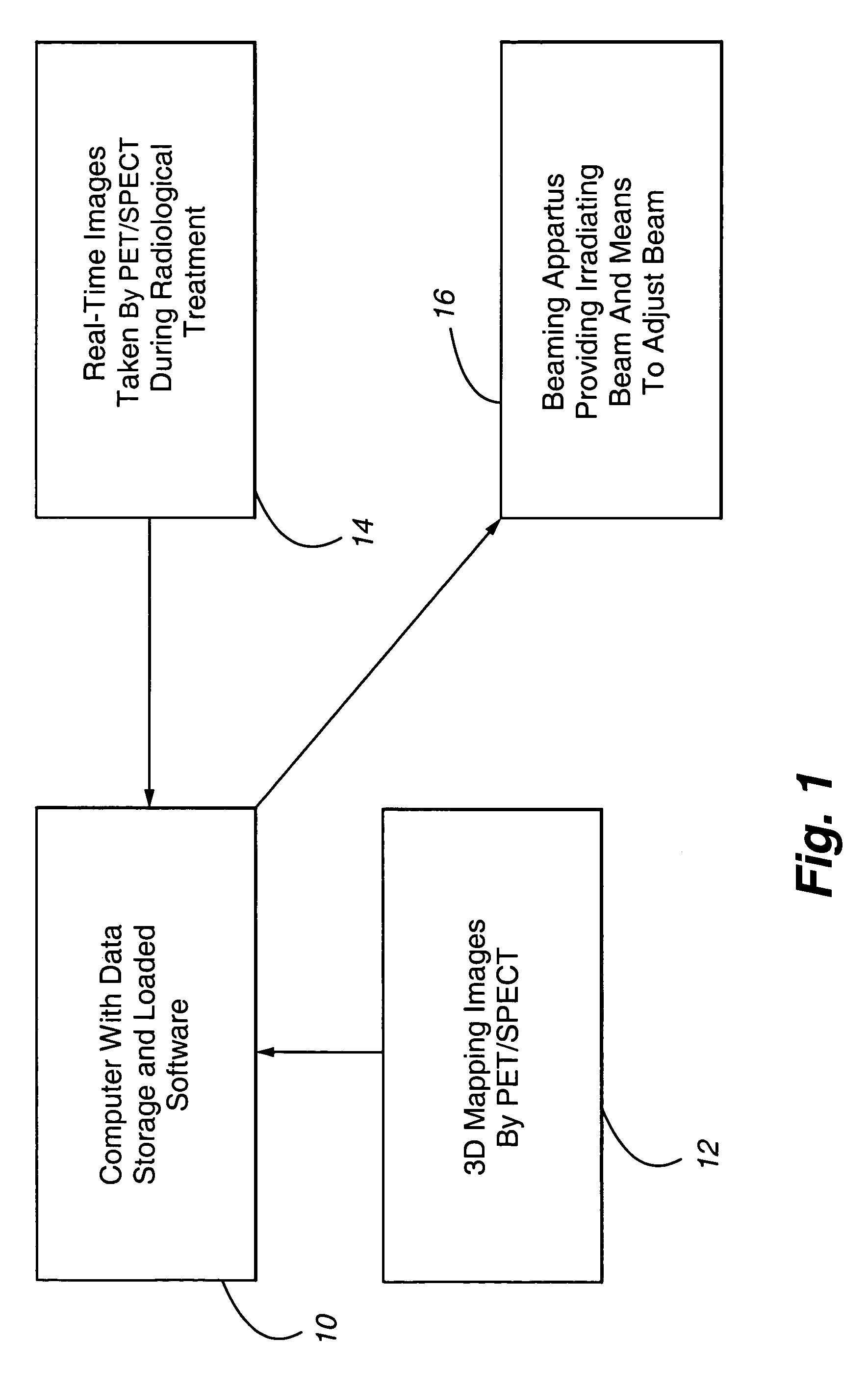
Figure 1
Abstract
Description
CROSS REFERENCE TO RELATED APPLICATIONS
[0001] This application is a continuation-in-part of application Ser. No. 10 / 176,558, filed on Jun. 21, 2002 now U.S. Pat. No. 6,750,037, entitled “Method of Cancer Screening Primarily Utilizing Non-Invasive Cell Collection, Fluorescence Detection Techniques, and Radio Tracing Detection Techniques”, the disclosure of which is hereby incorporated by reference herein.FIELD OF THE INVENTION
[0002] This invention relates to cancer screening and cancer treatment, and more particularly, to the use of metalloporphyrins for subsequent optimization of radiosurgery and / or radiotherapy.BACKGROUND OF THE INVENTION
[0003] There are a number of prior art methods and apparatuses which are used in the detection and treatment of cancer. Fluorescent markers have been used to help identify cancerous tissue within a patient. Radio tracers or markers have also been used in the detection and treatment of cancer.
[0004] U.S. Pat. No. 5,391,547 discloses a method of using po...
Examples
Embodiment Construction
[0030]In order to execute the method and apparatus of the present invention, a system is provided for delivering a collimated ionizing beam of radiation to a targeted area of tissue. Examples of such systems to support the present invention are disclosed in the U.S. Pat. Nos. 5,427,097 and 5,207,223.
[0031]Referring to FIG. 1, a representative system to achieve the present invention includes a computer 10 with data storage capability that is capable of executing instructions from software loaded within the computer. The computer can store and manipulate 3-dimensional mapping data images 12 of a patient being treated. The 3-dimensional mapping is typically stored in digital form, and is loaded in the computer 10 for later comparison purposes. As mentioned above, the 3-dimensional mapping is preferably achieved by SPECT or PET scanning following administration of a metaloporphyrin to a patient. A beaming apparatus 16 is provided which, when activated, emits a collimated surgical ionizi...