
Medicinal agent for inhibiting metastasis of malignant tumor
A malignant tumor and drug technology, which is applied in the direction of antineoplastic drugs, drug combinations, and pharmaceutical formulations, can solve the problems of hindering microvein adhesion, unclear role of physiological tumor metastasis, and great disparity.
Patent Information
- Authority / Receiving Office
- CN · China
- Patent Type
- Applications(China)
- Current Assignee / Owner
- Publication Date
- 2013-11-20
- Estimated Expiration
- Not applicable · inactive patent
Smart Images
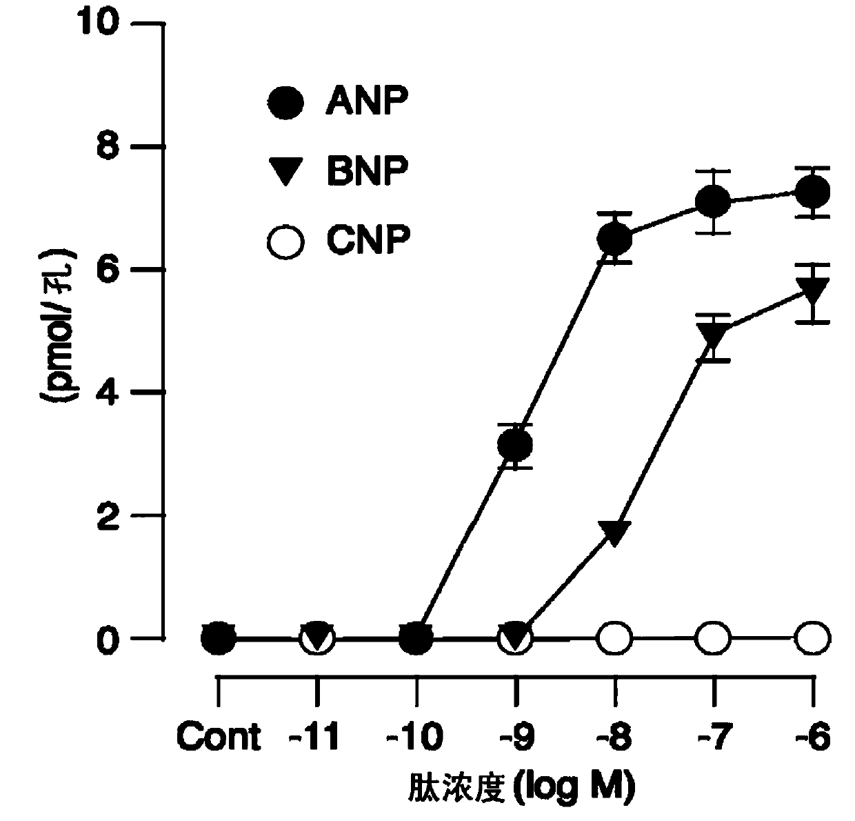
Figure 1 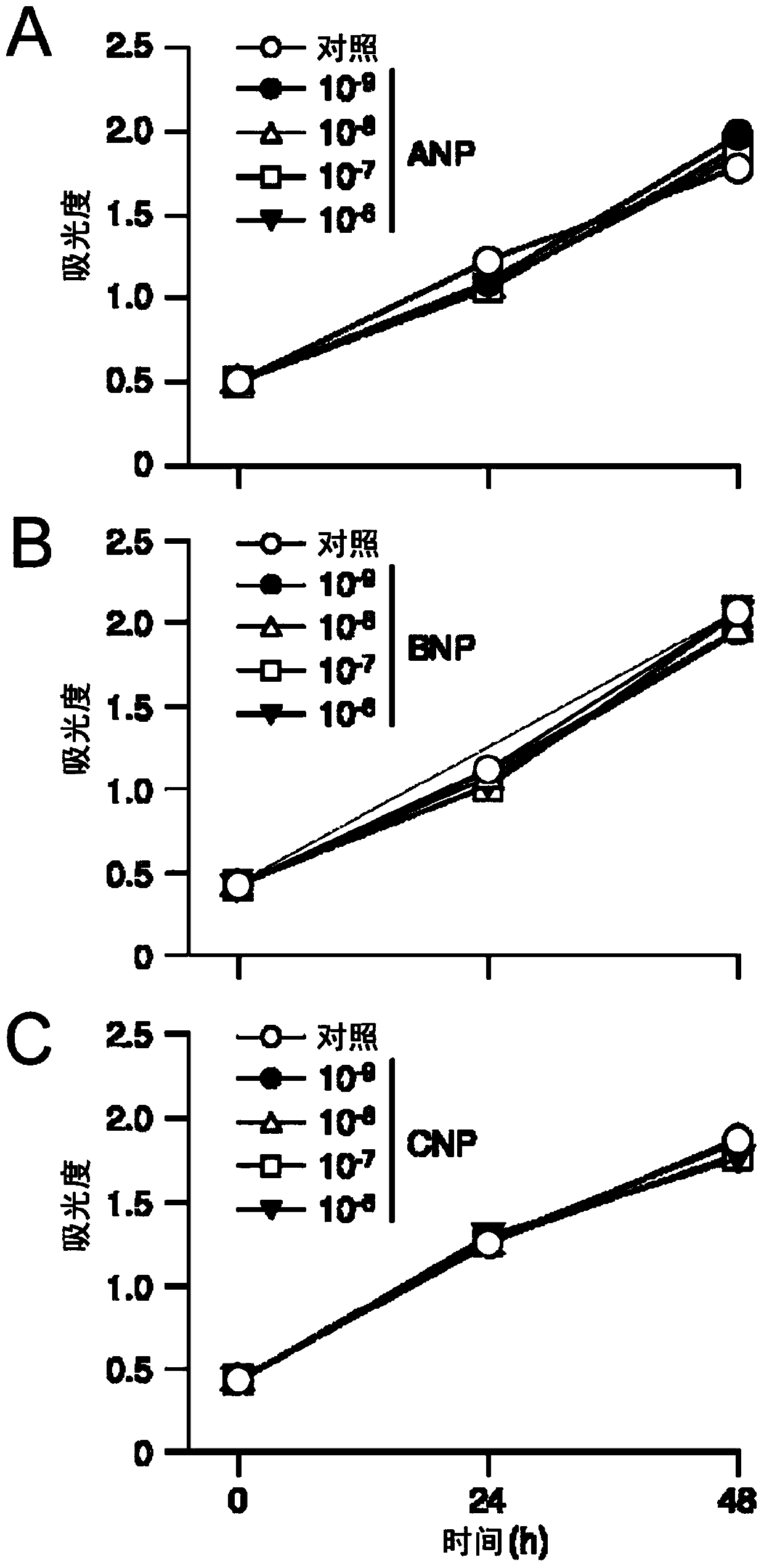
Figure 2 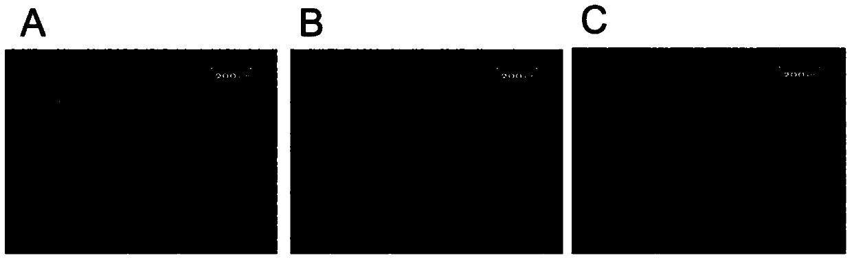
Figure 3
Abstract
Description
Technical field
[0001] The present invention relates to the metastasis of malignant tumors including cancer using vascular endothelial intracellular cGMP enhancers represented by natriuretic peptide receptors GC-A and GC-B agonists as active ingredients Pharmaceutical compositions for inhibition. Background technique
[0002] Malignant tumors represented by cancer are diseases based on the abnormal proliferation of cells. The biggest feature of malignant tumors is infiltration into surrounding tissues and metastasis to other organs. It has been known since ancient times that many of the causes of death in patients with malignant tumors are not the increase in the primary focus, but the multiple organ failure accompanied by the distant metastasis of tumor cells. The control of malignant tumor metastasis has not been achieved in recent years, and it is the most common cause of cancer treatment. One of the most important topics.
[0003] The metastasis of epithelial malignant tumors...
Examples
Embodiment 1
[0175] Changes in intracellular cGMP levels caused by natriuretic peptide stimulation on cancer cells
[0176] Divide A549 cells into 4×10 4 Cells / wells are added to a 24-well plate for culture, and the next day is replaced with DMEM without FCS. Add hANP, hBNP and hCNP solutions to the culture medium (final concentration: from 1×10 -11 M to 1×10 -6 M 10-fold dilution series, the control is the same amount of normal saline), 10 minutes later, 400 μL of cooled 70% ethanol (containing 0.1N hydrochloric acid) is added, and ultrasonic treatment is immediately performed. The treated solution was freeze-dried, and the cGMP level was measured using Cyclic GMP Assay kit (manufactured by Yamasa Corp.). The measurement results of cGMP levels are shown in figure 1 in.
[0177] HANP and hBNP, which are GC-A agonists, increase the intracellular cGMP of A549 cells in a concentration-dependent manner. On the other hand, in hCNP, which is a CG-B receptor agonist, which does not bind to the GC-...
Embodiment 2
[0178] Inhibitory effect of natriuretic peptide on the proliferation of A549 cells
[0179] Divide A549 cells into 4×10 4 Cells / wells are added to a 24-well plate for culture, and the next day is replaced with DMEM without FCS. Add hANP, hBNP and hCNP solutions to the culture medium (final concentration: from 1×10 -9 M to 1×10 -6 M 10-fold dilution series, the control is the same amount of physiological saline), cell proliferation analysis was performed 24 and 48 hours later using viable cell count reagent SF (manufactured by Nacalai Tesque). Show the result in figure 2 in.
[0180] Regarding the number of A549 cells until 48 hours later, even if it is up to 1×10 -6 Compared with the control, the concentration of M, hANP, hBNP and hCNP were basically unchanged. According to a report by Vesely et al., ANP has the effect of inhibiting the proliferation of various cancer cells derived from pancreatic cancer, breast cancer, etc., but in the test system of the example of the present ...
Embodiment 3
[0181] The effect of hANP on EMT induced by TGF-β1
[0182] Divide A549 cells into 1×10 5 Cells / wells were added to a 6-well plate for culture, the next day, the culture medium was replaced with DMEM without FBS, and the 24-hour culture medium was used for the following experiments.
[0183] The ANP group (addition of ANP solution (final concentration: 1μM)) and control group (addition of the same amount of physiological saline) were made, and the final concentration reached 0.125, 0.25, 0.5, 1.0 and 2.0ng / ml after 2 hours of addition. Method to add TGF-β1, 24 hours later, observe the morphological changes of the cells under the microscope, and take photos ( image 3 ).
[0184] After that, total RNA was recovered using total RNA isolation reagent (Invitrogen), cDNA was synthesized using reverse transcriptase, and E-cadherin, N-cadherin, VEGF-A, PDGF-B were measured by quantitative RT-PCR method , TNF-α and IL-6 mRNA levels. The results of mRNA level measurement are shown in Figu...