
Digital microscope and image identification method
A digital microscope and image technology, applied in the field of digital microscope, can solve problems such as poor image quality and result error, and achieve the effect of clear presentation, meeting medical needs, and accurate component identification.
Patent Information
- Authority / Receiving Office
- CN · China
- Current Assignee / Owner
- Publication Date
- 2014-12-31
- Estimated Expiration
- Not applicable · inactive patent
Smart Images
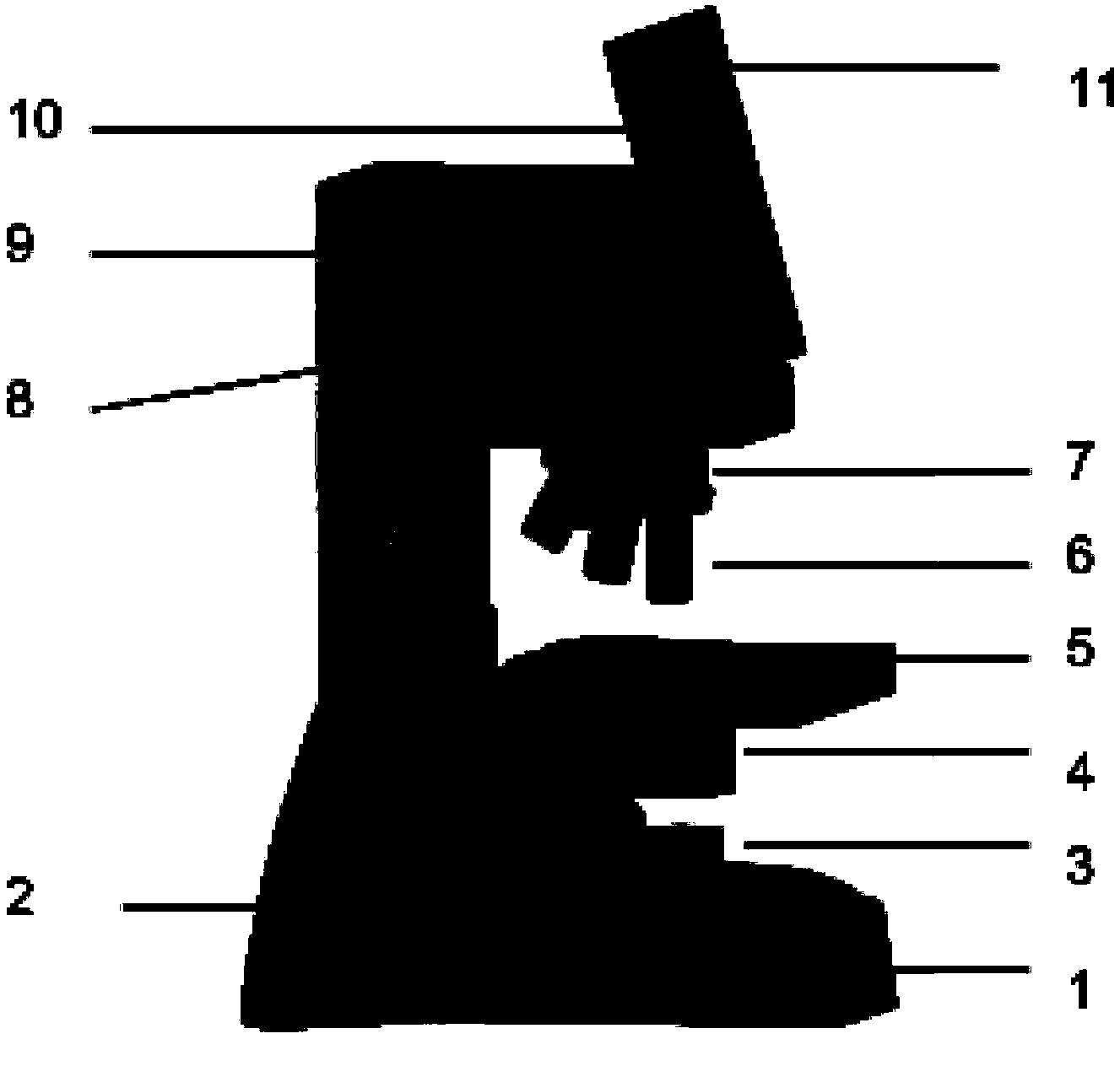
Figure 1 
Figure 2 
Figure 3
Abstract
Description
technical field
[0001] The invention relates to a digital microscope for urine sediment analysis, in particular to a digital microscope integrated with a display. Background technique
[0002] Urine is one of the important excretions in the body, and it is a window to reflect the pathological changes of kidney organs. As an indispensable and important part of complete urinalysis, urine sediment examination is of great significance for the diagnosis, localization, identification, prognosis judgment, drug treatment monitoring and health screening of diseases such as the urinary system. Urinary sediment refers to the shaped components in urine, which is the sediment formed after centrifugation of the original urine. It is a combination of quality and quantity of the formed components of urine, including cells, tubes, crystals, bacteria, sperm, etc.
[0003] Traditional manual microscope examination of urine sediment is not only labor-intensive and easy to pollute the environme...
Examples
Embodiment 1
[0037] This embodiment provides a digital microscope for urine sediment detection, its structure is as follows figure 1 shown, including:
[0038] An upright optical microscope device, which includes from bottom to top: microscope support 1, focusing device 2, lighting device 3, condenser lens 4, sample stage 5, objective lens 6, mirror changer seat 7, lens barrel lens (not shown in the figure) shown), in which the microscope stand 1 is used to support other components of the digital microscope, the illuminating device 3 is located under the sample stage 5, and the condenser lens 4 is located between the illuminating device 3 and the sample stage 5, and is used to convert the light emitted by the illuminating device 3 into Converging to the sample stage 5 to illuminate the urine sediment sample carried therein, the mirror changing rotary seat 7 is located above the sample stage 5, and a plurality of objective lenses 6 with different magnifications are installed on it, and the ...