
Automatic retinal vessel segmentation method based on non-fluorescent fundus images
A technology for automatic segmentation of retinal blood vessels, which is applied in the field of image processing, can solve the problems of low segmentation efficiency, low segmentation accuracy of tiny blood vessels, and easy adhesion, etc., and achieve the effects of suppressing uneven illumination, improving segmentation effects, and increasing contrast
Patent Information
- Authority / Receiving Office
- CN · China
- Patent Type
- Patents(China)
- Current Assignee / Owner
- Publication Date
- 2019-03-29
Smart Images
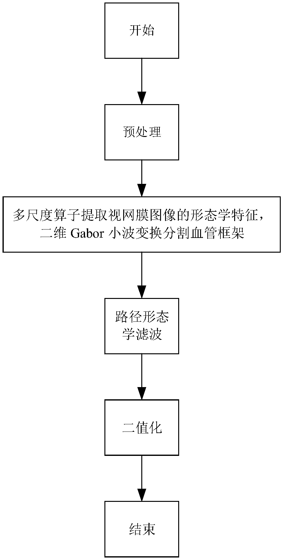
Figure 1 
Figure 2 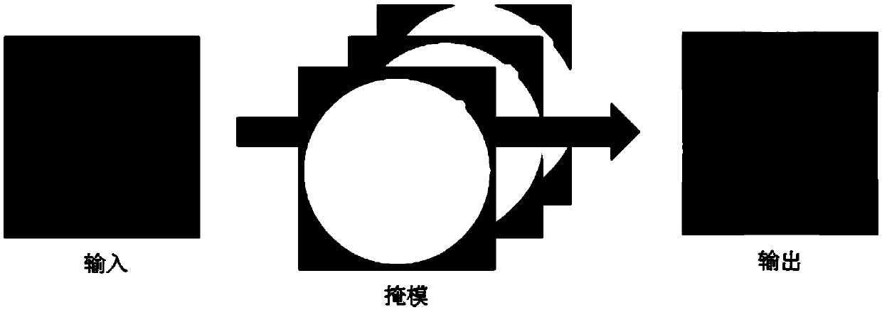
Figure 3
Abstract
Description
technical field
[0001] The invention relates to the technical field of image processing, in particular to an automatic retinal blood vessel segmentation method based on non-fluorescence fundus images. Background technique
[0002] In 1989, Chaudhuri et al. published the first two-dimensional matched filter method in "Detection of blood vessels in retinal images using two-dimensional matched filters". This method assumes that blood vessels are some equal-width line segments, and the width of blood vessels is between 2- 10 pixels, and the gray distribution of the blood vessel cross-section can be approximated by a Gaussian curve. Based on this, 12 Gaussian templates in different directions are constructed to perform matching filtering on the image, and the maximum response result is output as the filtering result. At that time, this method achieved excellent enhancement effects on blood vessels in retinal images, but the disadvantage was that when the image was doped with nois...
Examples
Embodiment
[0047] This embodiment includes the following steps:
[0048] The first step is to preprocess the image to enhance the characteristics of blood vessels and weaken the background noise.
[0049] The preprocessing includes contrast enhancement and retinal border growth.
[0050] Contrast enhancement is mainly based on a contrast-limited adaptive histogram equalization algorithm. The R channel is overexposed and the contrast is low; the brightness of the B channel is low, and blood vessels are difficult to identify; compared with the R and B channels, the contrast between the blood vessels and the background of the G channel image is the highest, and the noise is less. Therefore, we choose the G channel image for subsequent processing. The present invention uses the CLAHE algorithm to improve the local contrast of the G channel image, expecting to present more image details. Compared with the common adaptive histogram equalization method, the characteristic of CLAHE lies in it...