
Automatic segmentation method of breast tumor based on 3D MRI image
A technology for automatic segmentation of breast tumors, applied in the field of medical image processing, to achieve the effect of saving time, reducing workload, and high segmentation accuracy
Patent Information
- Authority / Receiving Office
- CN · China
- Patent Type
- Patents(China)
- Current Assignee / Owner
- Publication Date
- 2020-12-04
Smart Images
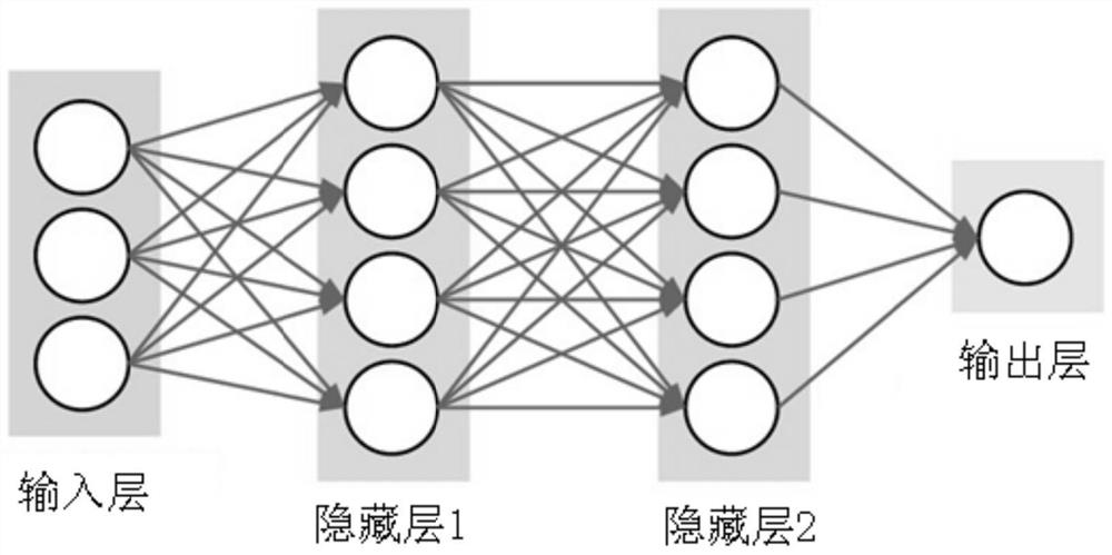
Figure 1 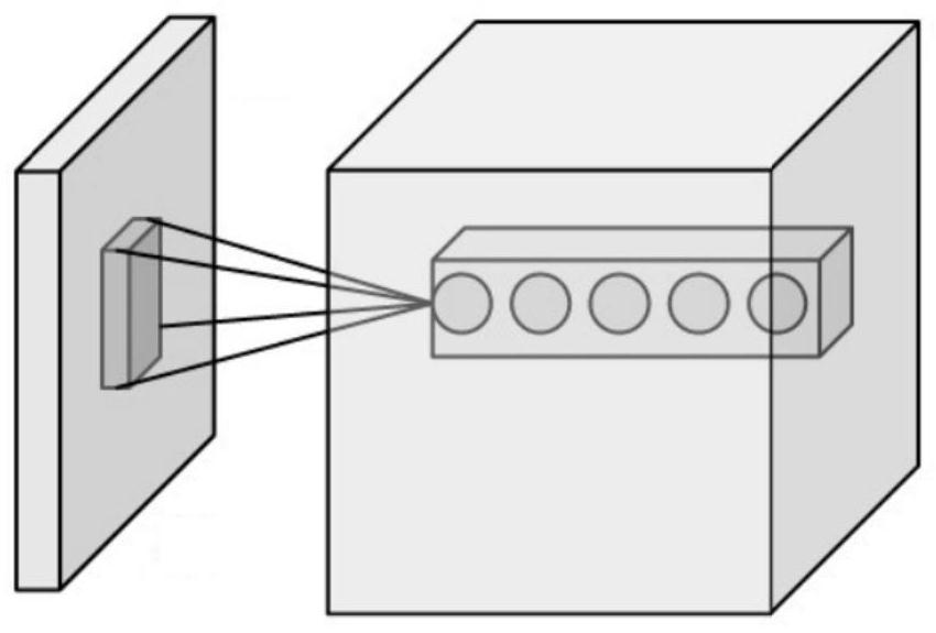
Figure 2 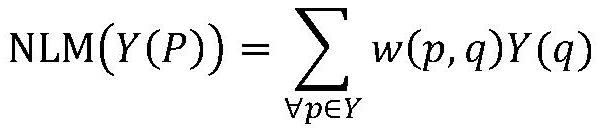
Figure 3
Abstract
Description
technical field
[0001] The invention belongs to the field of medical image processing, in particular to a method for automatically segmenting breast tumors based on three-dimensional MRI images. Background technique
[0002] Breast cancer is the most common malignant tumor in women, and its mortality rate ranks first among female malignant tumors in the world. At present, early diagnosis and timely treatment are the most effective measures to deal with breast cancer. Medical imaging methods, such as X-rays, magnetic resonance imaging (MRI), and ultrasound detection, are currently the most important means of detecting and diagnosing breast cancer. Among them, MRI can better distinguish various tissues and provide sufficient reference information for doctors. However, in general, doctors need to manually segment breast tumors, which takes a lot of time and effort, and the diagnostic accuracy is affected by the professional ability, experience and subjective factors of doctor...
Examples
Embodiment Construction
[0012] In order to make the technical problems, technical solutions and beneficial effects to be solved by the present invention clearer, the present invention will be further described in detail below in conjunction with the embodiments. It should be understood that the specific embodiments described here are only used to explain the present invention, not to limit the present invention.
[0013] Embodiments of the present invention provide a method for automatically segmenting breast tumors based on three-dimensional MRI images, comprising the following steps:
[0014] S01. Image preprocessing: provide an initial MRI image, and use a non-local average filter to preprocess the initial MRI image;
[0015] S02. Breast tumor location: build a multi-layer processing model for the training set, use convolutional neural network to perform hierarchical abstraction on the features of the training object, automatically extract segmentation features, and output the probability distribu...