
Isolated culture and differentiation method for testicular mesenchymal stem cells
A technology for separation and culture of testicular mesenchyme, applied in the biological field, can solve the problems of lack of methods for mesenchymal stem cell separation, culture and differentiation, and achieve the effect of simple, effective and highly controllable methods
Patent Information
- Authority / Receiving Office
- CN · China
- Patent Type
- Applications(China)
- Current Assignee / Owner
- Publication Date
- 2017-05-31
- Estimated Expiration
- Not applicable · inactive patent
Smart Images
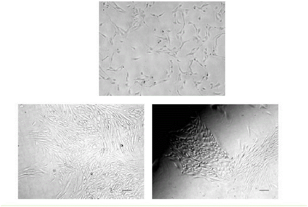
Figure 1 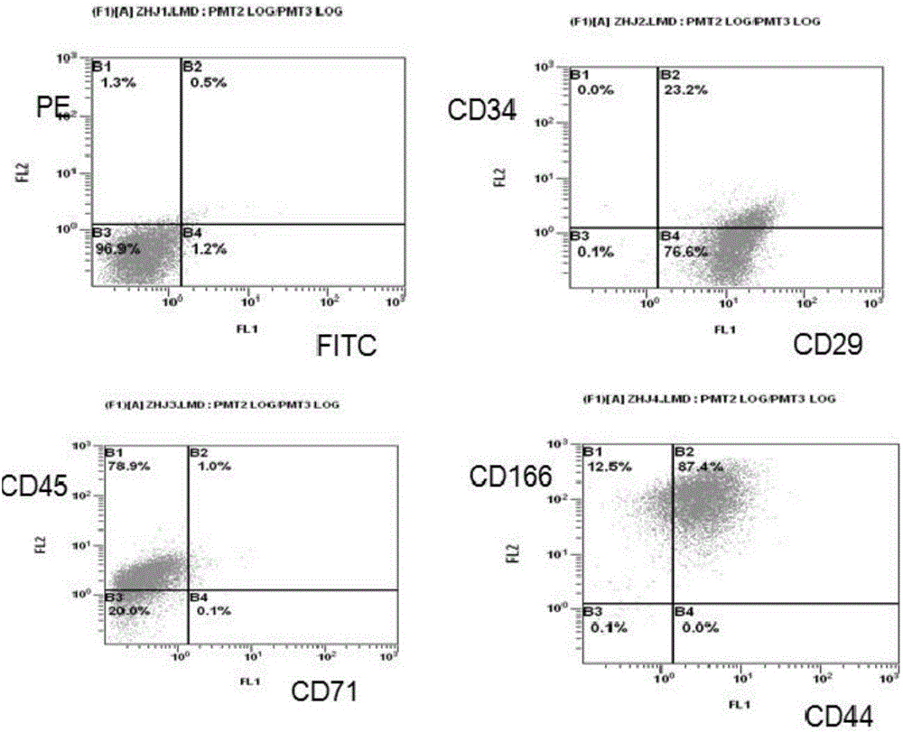
Figure 2 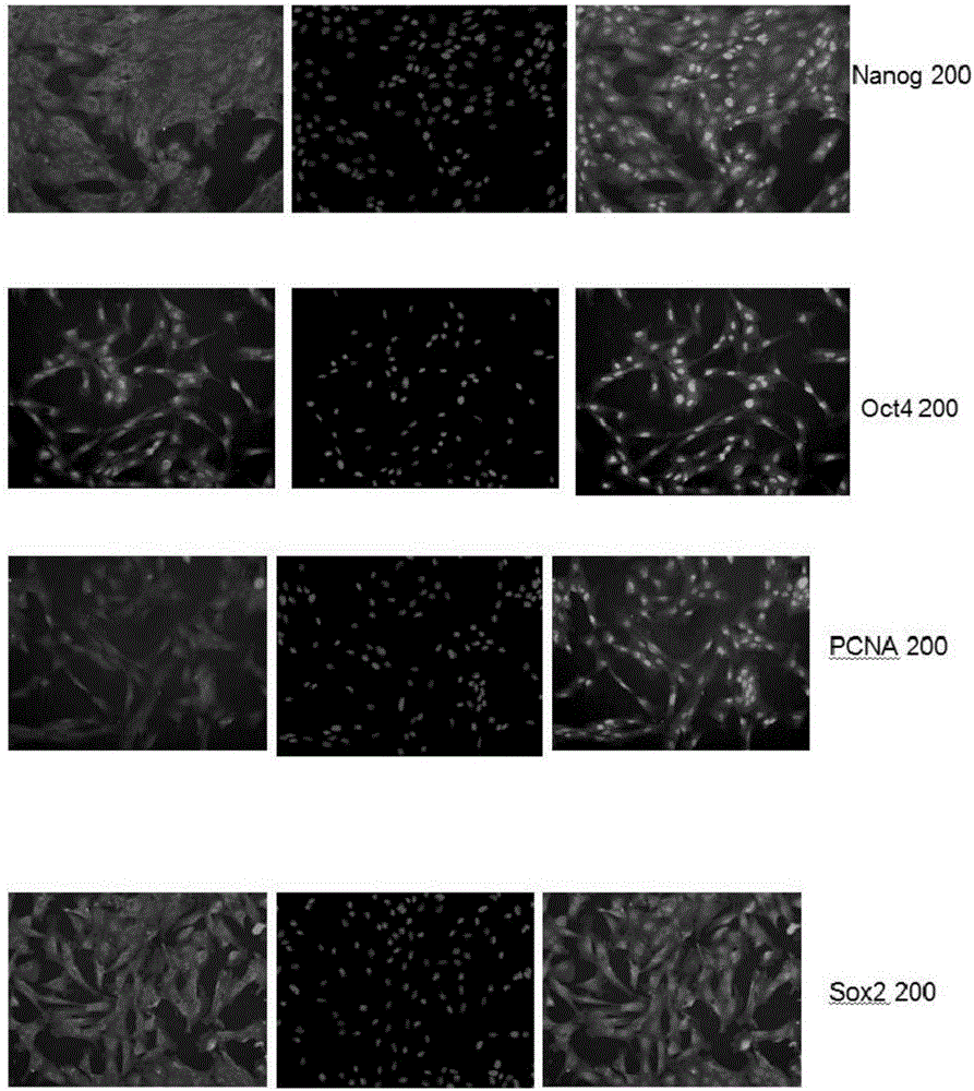
Figure 3
Abstract
Description
technical field
[0001] The invention relates to the field of biotechnology, in particular to a method for separating, culturing and differentiating testicular mesenchymal stem cells. Background technique
[0002] At present, stem cell-related technologies and achievements are constantly emerging, and stem cell research is experiencing a vigorous research boom. A variety of stem cell research, including embryonic stem cells, induced pluripotent stem cells and adult stem cells, has made great progress. Adult stem cells have gained more favor in clinical applications because of their low immunogenicity and high differentiation similarity with somatic cells. Bone marrow mesenchymal stem cells entered the field of vision of researchers more than 20 years ago, and adipose mesenchymal stem cells have also achieved fruitful results and considerable progress this year. Animal testes contain a large number of stem cells. In addition to male reproductive stem cells, testicular mesenc...
Examples
Embodiment
[0023] A method for isolating, culturing and differentiating testicular mesenchymal stem cells, comprising the following steps:
[0024] Step 1: Digest the obtained testicular tissue with trypsin and DNase, then use an equal volume of DMEM / F12 and 10% FBS solution to terminate the enzyme digestion, centrifuge at room temperature at 300g*5min, discard the supernatant; use 5mL DMEM / F12 and 10% FBS solution to resuspend the cells, centrifuge at 300g*5min at room temperature, discard the supernatant, and then culture and expand;
[0025] Step 2, culturing the P4 generation cells to form embryoid bodies;
[0026] In step 3, the embryoid bodies are inoculated and differentiated.
Embodiment 2
[0028] A method for isolating, culturing and differentiating testicular mesenchymal stem cells, comprising the following steps:
[0029] Step 1, the method of separating the obtained testicular tissue in vitro is: digest the obtained testicular tissue with 2mg / mL collagenase, 20μg / mL DNase and 2mg / mL decomposing enzyme for 20min, and then use 1mg / mL trypsin and 10μg / mL Digest with mL DNase for 10 minutes, then stop the enzyme digestion with an equal volume of DMEM / F12 plus 10% FBS solution, centrifuge at 300g*5min at room temperature, discard the supernatant; resuspend the cells with 5mL DMEM / F12 plus 10% FBS solution, at room temperature 300g * Centrifuge for 5 minutes, discard the supernatant, and then culture and amplify;
[0030] Step 2: Digest the well-cultured P4 cells in step 1 with 0.25% trypsin, centrifuge at 300g*5min, resuspend in PBS buffer, and centrifuge at 300g*5min; resuspend with DMEM / F12 plus 10% FBS Cells were counted on a hemocytometer, and the cell densit...
Embodiment 3
[0033] A method for isolating, culturing and differentiating testicular mesenchymal stem cells, comprising the following steps:
[0034] Step 1, the method of separating the obtained testicular tissue in vitro is: digest the obtained testicular tissue with 2mg / mL collagenase, 20μg / mL DNase and 2mg / mL decomposing enzyme for 20min, and then use 1mg / mL trypsin and 10μg / mL Digest with mL DNase for 10 minutes, then stop the enzyme digestion with an equal volume of DMEM / F12 plus 10% FBS solution, centrifuge at 300g*5min at room temperature, discard the supernatant; resuspend the cells with 5mL DMEM / F12 plus 10% FBS solution, at room temperature 300g * Centrifuge for 5 minutes, discard the supernatant, and then culture and amplify;
[0035] Step 2: Digest the well-cultured P4 cells in step 1 with 0.25% trypsin, centrifuge at 300g*5min, resuspend in PBS buffer, and centrifuge at 300g*5min; resuspend with DMEM / F12 plus 10% FBS Cells were counted on a hemocytometer, and the cell densit...