
MRT imaging on the basis of conventional PPA reconstruction methods
a reconstruction method and imaging technology, applied in the field of magnetic resonance tomography (mrt), can solve the problems of image artifacts and residual apparatus-dependent artifacts, and achieve the effect of improving the resolution and improving the ppa imaging of slice images
Patent Information
- Authority / Receiving Office
- US · United States
- Current Assignee / Owner
- Publication Date
- 2006-11-23
- Estimated Expiration
- Not applicable · inactive patent
Smart Images
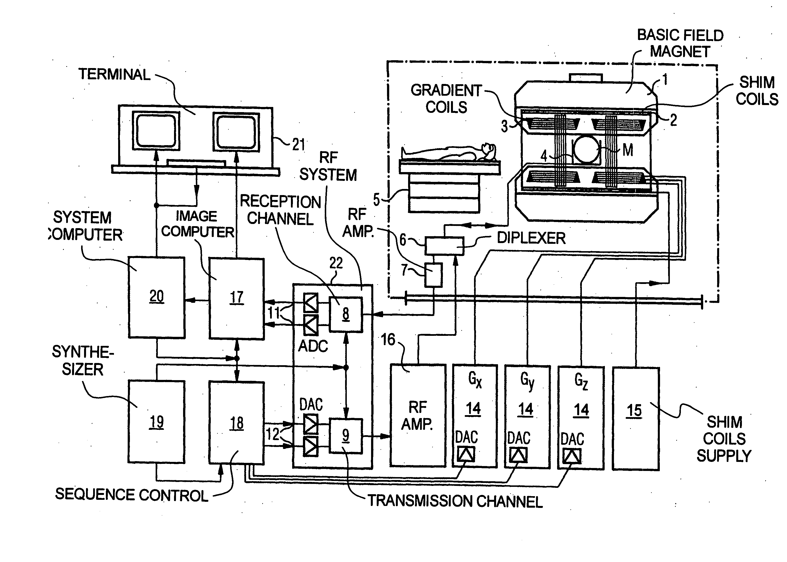
Figure 1 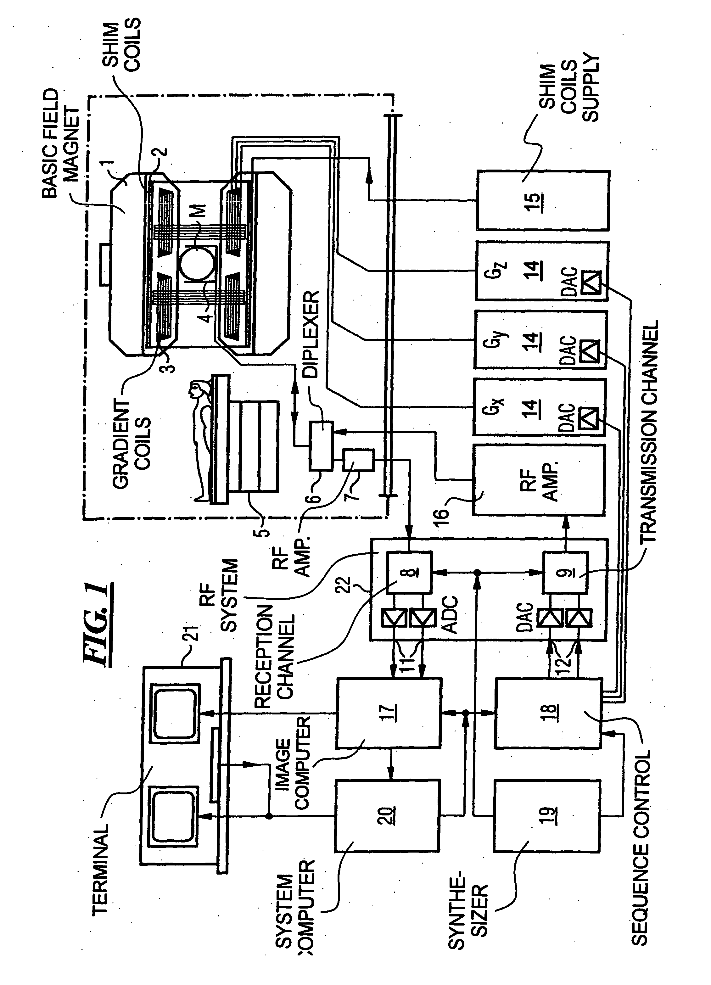
Figure 2 
Figure 3
Abstract
Description
BACKGROUND OF THE INVENTION
[0001] 1. Field of the Invention
[0002] The present invention generally concerns magnetic resonance tomography (MRT) as employed in medicine for examination of patients. The present invention concerns a method as well as an MRT system for implementation of the method that enable the acquisition of reduced-artifact or improved-resolution slice images that are displaced relative to one another in the z-gradient direction.
[0003] 2. Description of the Prior Art
[0004] MRT is based on the physical phenomenon of nuclear magnetic resonance and has been successfully used as an imaging method for over 15 years in medicine and biophysics. In this examination modality, the subject is exposed to a strong, constant magnetic field. The nuclear spins of the atoms in the subject, which were previously randomly oriented, thereby align.
[0005] Radio-frequency energy can now excite these “ordered” nuclear spins to a specific oscillation. In MRT, this oscillation generates ...
Examples
Embodiment Construction
[0041]FIG. 1 is a schematic illustration of a magnetic resonance imaging or magnetic resonance tomography apparatus for generating a magnetic resonance image of a subject according to the present invention. The design of the magnetic resonance tomography apparatus thereby corresponds that of a conventional magnetic tomography apparatus, with the differences identified below. A basic field magnet 1 generates a temporally-constant strong magnetic field for polarization or alignment of the nuclear spins in the examination region of a subject such as, for example, of a part of a human body to be examined. The high homogeneity of the basic magnetic field necessary for the magnetic resonance measurement is defined in a spherical measurement volume V in which the parts of the human body to be examined are introduced. To support the homogeneity requirements, and in particular for elimination of temporally invariable influences, shim plates made of ferromagnetic material are mounted at a sui...