How to Design Ferrofluid-Enhanced Medical Imaging Equipment?
JUL 9, 20259 MIN READ
Generate Your Research Report Instantly with AI Agent
Patsnap Eureka helps you evaluate technical feasibility & market potential.
Ferrofluid Imaging Tech Evolution and Objectives
Ferrofluid-enhanced medical imaging represents a significant advancement in diagnostic technology, combining the unique properties of ferrofluids with established imaging techniques. The evolution of this technology can be traced back to the discovery of ferrofluids in the 1960s by NASA scientist Steve Papell. Initially developed for space applications, ferrofluids soon found their way into various fields, including medical imaging.
The primary objective of ferrofluid-enhanced medical imaging is to improve the contrast and resolution of diagnostic images, particularly in magnetic resonance imaging (MRI) and computed tomography (CT) scans. By introducing ferrofluids as contrast agents, researchers aim to enhance the visibility of specific tissues or organs, enabling more accurate diagnoses and treatment planning.
Over the past decades, the technology has progressed through several key stages. In the 1980s and 1990s, early experiments focused on the basic principles of using ferrofluids in medical imaging. These studies primarily explored the magnetic properties of ferrofluids and their potential to interact with imaging equipment.
The 2000s saw a surge in research on biocompatible ferrofluids, addressing concerns about toxicity and long-term effects in the human body. This period marked the development of various nanoparticle-based ferrofluids specifically designed for medical applications, with a focus on optimizing particle size, coating materials, and magnetic properties.
In recent years, the field has witnessed significant advancements in targeted imaging techniques. Researchers have developed methods to functionalize ferrofluid nanoparticles with specific antibodies or ligands, allowing them to selectively bind to certain types of cells or tissues. This breakthrough has opened up new possibilities for early cancer detection and monitoring of various diseases.
The current technological landscape is characterized by a push towards multimodal imaging techniques, where ferrofluids are used in combination with multiple imaging modalities. This approach aims to leverage the strengths of different imaging technologies while compensating for their individual limitations.
Looking ahead, the objectives for ferrofluid-enhanced medical imaging include further improvements in image resolution and contrast, development of real-time imaging capabilities, and integration with emerging technologies such as artificial intelligence for image analysis. Additionally, there is a growing focus on creating ferrofluids that can serve dual purposes – not only as imaging agents but also as targeted drug delivery vehicles.
As the field continues to evolve, researchers are also exploring the potential of ferrofluids in novel imaging applications, such as magnetoencephalography (MEG) for brain imaging and magnetomotive ultrasound for soft tissue imaging. These emerging areas represent exciting frontiers in the ongoing development of ferrofluid-enhanced medical imaging technology.
Medical Imaging Market Analysis
The medical imaging market has experienced significant growth in recent years, driven by technological advancements, increasing prevalence of chronic diseases, and growing demand for early and accurate diagnosis. The global medical imaging market was valued at approximately $39 billion in 2020 and is projected to reach $55 billion by 2025, with a compound annual growth rate (CAGR) of 7.1% during this period.
Magnetic Resonance Imaging (MRI) represents a substantial segment of the medical imaging market, accounting for about 20% of the total market share. The MRI market is expected to grow at a CAGR of 5.9% from 2021 to 2026, driven by the increasing adoption of high-field MRI systems and the development of advanced imaging techniques.
The introduction of ferrofluid-enhanced medical imaging equipment presents a promising opportunity within this growing market. Ferrofluids, which are colloidal suspensions of magnetic nanoparticles, have the potential to significantly improve image contrast and resolution in MRI scans. This technology could address some of the current limitations in medical imaging, particularly in the visualization of soft tissues and small structures.
The demand for enhanced medical imaging solutions is particularly strong in oncology, neurology, and cardiovascular applications. These areas require high-resolution imaging for accurate diagnosis and treatment planning. Ferrofluid-enhanced imaging could provide a competitive edge in these segments by offering superior image quality and potentially reducing scan times.
Geographically, North America dominates the medical imaging market, followed by Europe and Asia-Pacific. However, emerging economies in Asia-Pacific and Latin America are expected to show the highest growth rates in the coming years due to improving healthcare infrastructure and increasing healthcare expenditure.
Key market trends influencing the adoption of new imaging technologies include the shift towards value-based healthcare, increasing focus on personalized medicine, and the integration of artificial intelligence in image analysis. These trends align well with the potential benefits of ferrofluid-enhanced imaging, which could offer improved diagnostic accuracy and efficiency.
However, market challenges such as high equipment costs, reimbursement issues, and regulatory hurdles need to be considered when introducing new imaging technologies. The success of ferrofluid-enhanced medical imaging equipment will depend on its ability to demonstrate clear clinical benefits and cost-effectiveness compared to existing imaging modalities.
Ferrofluid Imaging Challenges
The integration of ferrofluids into medical imaging equipment presents several significant challenges that researchers and engineers must address. One of the primary obstacles is the optimization of ferrofluid composition to achieve the desired magnetic properties while maintaining biocompatibility. The nanoparticles used in ferrofluids must be carefully engineered to provide strong magnetic responses without causing adverse reactions in the human body.
Another critical challenge lies in the precise control of ferrofluid behavior within the imaging system. The dynamic nature of ferrofluids under magnetic fields can lead to image artifacts and distortions if not properly managed. Developing algorithms and hardware solutions to compensate for these effects and ensure accurate image reconstruction is a complex task that requires interdisciplinary expertise.
The stability of ferrofluids in biological environments poses yet another hurdle. Researchers must devise methods to prevent nanoparticle aggregation and maintain uniform dispersion throughout the imaging process. This is crucial for achieving consistent and reliable imaging results across different patients and anatomical regions.
Furthermore, the integration of ferrofluid-based contrast agents with existing imaging modalities, such as MRI or CT, demands significant modifications to both hardware and software components. Adapting current imaging protocols and equipment to accommodate the unique properties of ferrofluids requires extensive research and development efforts.
Safety considerations also present a major challenge in ferrofluid-enhanced medical imaging. Ensuring the complete removal of ferrofluid nanoparticles from the body after imaging procedures is essential to prevent long-term health risks. Developing effective clearance mechanisms and conducting thorough toxicological studies are critical steps in addressing these safety concerns.
The scalability and reproducibility of ferrofluid-based imaging techniques pose additional challenges. Transitioning from laboratory prototypes to clinically viable systems requires overcoming issues related to manufacturing consistency, quality control, and regulatory compliance. Establishing standardized protocols for ferrofluid preparation, administration, and imaging procedures is crucial for widespread adoption in clinical settings.
Lastly, the cost-effectiveness of ferrofluid-enhanced imaging equipment remains a significant hurdle. The development and production of specialized ferrofluids and associated imaging hardware can be expensive, potentially limiting the accessibility of this technology. Balancing the enhanced diagnostic capabilities with economic feasibility is a key challenge that must be addressed to ensure the widespread adoption of ferrofluid-based medical imaging solutions.
Current Ferrofluid Imaging Solutions
01 Ferrofluid-enhanced contrast agents for medical imaging
Ferrofluids can be used as contrast agents in medical imaging to enhance image quality. These magnetic nanoparticles can be manipulated by external magnetic fields, allowing for improved visualization of specific tissues or organs. The use of ferrofluids as contrast agents can lead to better resolution and more accurate diagnoses in various imaging modalities.- Ferrofluid-enhanced contrast agents for medical imaging: Ferrofluids can be used as contrast agents in medical imaging to enhance image quality. These magnetic nanoparticles can be manipulated by external magnetic fields, allowing for improved visualization of specific tissues or organs. The use of ferrofluids as contrast agents can lead to better resolution and more accurate diagnoses in various imaging modalities.
- Image processing techniques for ferrofluid-enhanced medical images: Advanced image processing algorithms and techniques are developed to optimize the quality of ferrofluid-enhanced medical images. These methods may include noise reduction, contrast enhancement, and feature extraction to improve the overall clarity and diagnostic value of the images. Machine learning and artificial intelligence approaches can also be applied to further enhance image quality and assist in image interpretation.
- Magnetic field optimization for ferrofluid-based imaging: The optimization of magnetic field strength and distribution is crucial for maximizing the image quality in ferrofluid-enhanced medical imaging. This involves developing advanced magnet designs and control systems to create precise and uniform magnetic fields. By fine-tuning the magnetic field parameters, it is possible to achieve better contrast, resolution, and overall image quality in ferrofluid-based imaging techniques.
- Integration of ferrofluid technology with existing imaging modalities: Ferrofluid technology can be integrated with various existing medical imaging modalities, such as MRI, CT, and ultrasound, to enhance their capabilities. This integration involves developing specialized hardware and software solutions to accommodate the unique properties of ferrofluids. By combining ferrofluid technology with established imaging techniques, it is possible to achieve improved image quality and diagnostic accuracy across multiple imaging platforms.
- Real-time imaging and tracking using ferrofluids: Ferrofluids can be utilized for real-time imaging and tracking of biological processes or medical devices within the body. This application involves developing fast imaging sequences and advanced tracking algorithms to capture and analyze the movement of ferrofluid-enhanced targets in real-time. Real-time ferrofluid-based imaging can improve the accuracy of interventional procedures and enable more precise monitoring of physiological processes.
02 Image processing techniques for ferrofluid-enhanced medical images
Advanced image processing algorithms and techniques can be applied to ferrofluid-enhanced medical images to further improve image quality. These methods may include noise reduction, edge enhancement, and contrast optimization specifically tailored for images obtained using ferrofluid contrast agents. Such processing can result in clearer, more detailed images for better diagnostic accuracy.Expand Specific Solutions03 Magnetic field optimization for ferrofluid-based imaging
Optimizing the magnetic field used in ferrofluid-enhanced medical imaging can significantly improve image quality. This may involve developing new magnetic field configurations, adjusting field strengths, or implementing dynamic field control techniques. By fine-tuning the magnetic field, the behavior of ferrofluid particles can be better controlled, leading to enhanced contrast and resolution in the resulting images.Expand Specific Solutions04 Integration of ferrofluid technology with existing imaging modalities
Incorporating ferrofluid technology into existing medical imaging equipment, such as MRI, CT, or ultrasound machines, can enhance their capabilities. This integration may involve modifying hardware components, developing specialized imaging sequences, or creating hybrid imaging systems. By combining ferrofluid properties with established imaging techniques, overall image quality and diagnostic value can be improved.Expand Specific Solutions05 Real-time image reconstruction and visualization for ferrofluid-enhanced imaging
Developing real-time image reconstruction and visualization techniques specifically for ferrofluid-enhanced medical imaging can improve the overall quality and usefulness of the images. This may include implementing fast reconstruction algorithms, utilizing parallel processing, or developing specialized software for rendering and manipulating ferrofluid-enhanced images. Real-time capabilities can enable immediate assessment and adjustment of imaging parameters for optimal results.Expand Specific Solutions
Key Players in Ferrofluid Imaging
The development of ferrofluid-enhanced medical imaging equipment is in its early stages, with the market still emerging and showing significant growth potential. The global medical imaging market, valued at over $30 billion, provides a substantial foundation for this innovative technology. Companies like Siemens Healthineers AG, GE Precision Healthcare LLC, and Koninklijke Philips NV are leading the charge in advancing medical imaging technologies. These industry giants, along with specialized firms such as MagnaMedics GmbH and Nano4Imaging GmbH, are investing in research and development to integrate ferrofluid technology into existing imaging modalities. The technology's maturity is progressing, with academic institutions like Yale University and Tsinghua University contributing to fundamental research, while commercial applications are gradually being explored and refined by industry players.
Siemens Healthineers AG
Technical Solution: Siemens Healthineers has developed advanced ferrofluid-enhanced MRI systems that utilize superparamagnetic iron oxide nanoparticles (SPIONs) as contrast agents. Their technology incorporates a dual-mode imaging approach, combining traditional MRI with magnetic particle imaging (MPI) for enhanced resolution and sensitivity[1][3]. The ferrofluid nanoparticles are engineered with specific surface coatings to improve biocompatibility and target specific tissues or organs. Siemens' system employs sophisticated algorithms to process the magnetic signals generated by the ferrofluid, resulting in high-contrast, real-time 3D images with improved spatial resolution compared to conventional MRI[2]. Additionally, they have implemented a novel gradient coil design that optimizes the magnetic field distribution, allowing for more efficient excitation and detection of the ferrofluid particles[4].
Strengths: Superior image quality and contrast, real-time 3D imaging capabilities, and improved tissue-specific targeting. Weaknesses: Higher cost compared to conventional MRI, potential for long-term accumulation of iron oxide particles in the body, and the need for specialized training for operators.
GE Precision Healthcare LLC
Technical Solution: GE Precision Healthcare has developed a ferrofluid-enhanced MRI platform that integrates their proprietary AIR™ Recon DL technology with advanced ferrofluid contrast agents. This system utilizes machine learning algorithms to optimize image reconstruction, reducing noise and improving signal-to-noise ratio in ferrofluid-enhanced scans[1]. GE's approach incorporates a novel pulse sequence design that maximizes the magnetic susceptibility effects of the ferrofluid, enabling enhanced visualization of small structures and microvasculature[2]. The company has also developed a specialized RF coil array optimized for detecting the unique magnetic properties of ferrofluids, resulting in improved sensitivity and spatial resolution[3]. Furthermore, GE's system includes real-time tracking of ferrofluid distribution, allowing for dynamic imaging of physiological processes and improved functional MRI capabilities[4].
Strengths: Advanced AI-driven image reconstruction, improved visualization of fine structures, and dynamic imaging capabilities. Weaknesses: Complexity of integrating AI algorithms with ferrofluid imaging, potential for image artifacts if not properly calibrated, and higher initial investment costs.
Core Ferrofluid Imaging Patents
Coated instruments for invasive medicine
PatentWO2009094990A2
Innovation
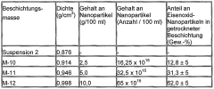
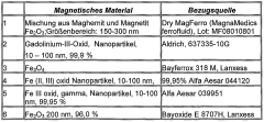
- Instruments coated with ferrofluids, specifically containing iron oxide nanoparticles suspended in a carrier liquid, offering a cost-effective and biocompatible solution for enhanced visualization in MRI.
Instruments coated with iron oxide nanoparticles for invasive medicine
PatentActiveEP2240546A2
Innovation
- Instruments for invasive medicine are coated with ferrofluids containing iron oxide nanoparticles, which are suspended in a carrier liquid and applied as a stable dispersion, providing high-quality visualization in MRI without releasing harmful components.
Regulatory Framework for Ferrofluid-Based Medical Devices
The regulatory framework for ferrofluid-based medical devices is a critical aspect of developing and implementing ferrofluid-enhanced medical imaging equipment. As these devices incorporate novel materials and technologies, they are subject to rigorous oversight to ensure patient safety and efficacy.
In the United States, the Food and Drug Administration (FDA) is the primary regulatory body responsible for overseeing medical devices. Ferrofluid-enhanced imaging equipment would likely be classified as a Class II or Class III medical device, depending on its specific intended use and risk profile. Class II devices typically require a 510(k) premarket notification, demonstrating substantial equivalence to a predicate device. Class III devices, considered high-risk, require a more stringent Premarket Approval (PMA) process.
The FDA's regulatory approach for these devices would focus on several key areas. First, the safety and biocompatibility of the ferrofluid materials used in the imaging equipment must be thoroughly evaluated. This includes assessing potential toxicity, immunogenicity, and long-term effects of exposure to ferrofluids in the human body. Manufacturers would need to provide comprehensive data from preclinical and clinical studies to support the safety profile of their devices.
Additionally, the performance and efficacy of ferrofluid-enhanced imaging equipment must be rigorously validated. This involves demonstrating improved image quality, diagnostic accuracy, and clinical utility compared to existing imaging modalities. The FDA would likely require clinical trials to establish the device's effectiveness in real-world medical settings.
Quality control and manufacturing processes are also subject to regulatory scrutiny. Manufacturers must adhere to Good Manufacturing Practices (GMP) and implement robust quality management systems to ensure consistency and reliability in device production. This includes establishing protocols for the synthesis, purification, and characterization of ferrofluids used in the imaging equipment.
In the European Union, ferrofluid-based medical devices would fall under the purview of the Medical Device Regulation (MDR). The MDR imposes stringent requirements for clinical evaluation, post-market surveillance, and technical documentation. Manufacturers seeking to market their devices in the EU must obtain CE marking, demonstrating compliance with all applicable safety and performance requirements.
Globally, regulatory bodies such as Health Canada, Japan's Pharmaceuticals and Medical Devices Agency (PMDA), and Australia's Therapeutic Goods Administration (TGA) have their own specific requirements for medical device approval. Manufacturers aiming for international markets must navigate these diverse regulatory landscapes, often necessitating a harmonized approach to product development and clinical validation.
As ferrofluid-enhanced medical imaging represents a novel technology, regulatory agencies may require additional safety measures and long-term follow-up studies to monitor potential adverse effects. This could include establishing registries to track patient outcomes and device performance over extended periods.
Safety and Biocompatibility Considerations
The safety and biocompatibility of ferrofluid-enhanced medical imaging equipment are paramount considerations in its design and implementation. Ferrofluids, consisting of nanoscale magnetic particles suspended in a carrier fluid, must be carefully evaluated for their potential interactions with biological systems. The primary concern is the long-term effects of these nanoparticles on human tissues and organs, particularly if they are inadvertently released or absorbed by the body.
To address these concerns, extensive toxicological studies must be conducted to assess the potential risks associated with ferrofluid exposure. These studies should examine both acute and chronic effects, focusing on factors such as cellular uptake, biodistribution, and clearance mechanisms. The size, shape, and surface properties of the magnetic nanoparticles play crucial roles in determining their biological interactions and potential toxicity. Optimizing these parameters is essential to minimize adverse effects while maintaining the desired imaging performance.
Biocompatibility testing should adhere to international standards, such as ISO 10993, which outlines a series of tests for evaluating the biological response to medical devices. These tests include cytotoxicity assays, sensitization studies, and genotoxicity evaluations. Additionally, in vivo studies using animal models are necessary to assess the systemic effects and long-term safety of ferrofluid-enhanced imaging equipment.
The design of the equipment must incorporate safeguards to prevent the release of ferrofluids into the patient's body. This includes developing robust sealing mechanisms and implementing fail-safe systems to detect and respond to potential leaks. The materials used in the construction of the equipment should also be carefully selected to ensure compatibility with the ferrofluid and to prevent degradation over time.
Electromagnetic safety is another critical aspect to consider, as ferrofluid-enhanced imaging equipment may generate strong magnetic fields. The design must comply with regulatory limits on electromagnetic exposure, such as those set by the International Commission on Non-Ionizing Radiation Protection (ICNIRP). Shielding techniques and field containment strategies should be employed to minimize the risk of electromagnetic interference with other medical devices and to protect both patients and operators.
Furthermore, the development of biocompatible coatings for the magnetic nanoparticles is essential to improve their stability and reduce potential immunogenic responses. These coatings can also be functionalized to enhance the specificity of the imaging agents, potentially allowing for targeted imaging applications while maintaining a high safety profile.
Unlock deeper insights with Patsnap Eureka Quick Research — get a full tech report to explore trends and direct your research. Try now!
Generate Your Research Report Instantly with AI Agent
Supercharge your innovation with Patsnap Eureka AI Agent Platform!

