Increased depth of field microscope and associated devices, systems, and methods

a microscope and depth-of-field technology, applied in the field of ophthalmic surgical microscopes, can solve the problems of reducing the amount of light passing through the microscope optics, reducing the photon flux or amount of light passing through the microscope, and expensive large objective lenses, etc., to improve the doctor's view of the surgical field.

US20160022133A1Active Publication Date: 2016-01-28ALCON INC
4 Cites 5 Cited by

Patent Information

Authority / Receiving Office
US · United States
Patent Type
Applications(United States)
Current Assignee / Owner
Publication Date
2016-01-28

Smart Images

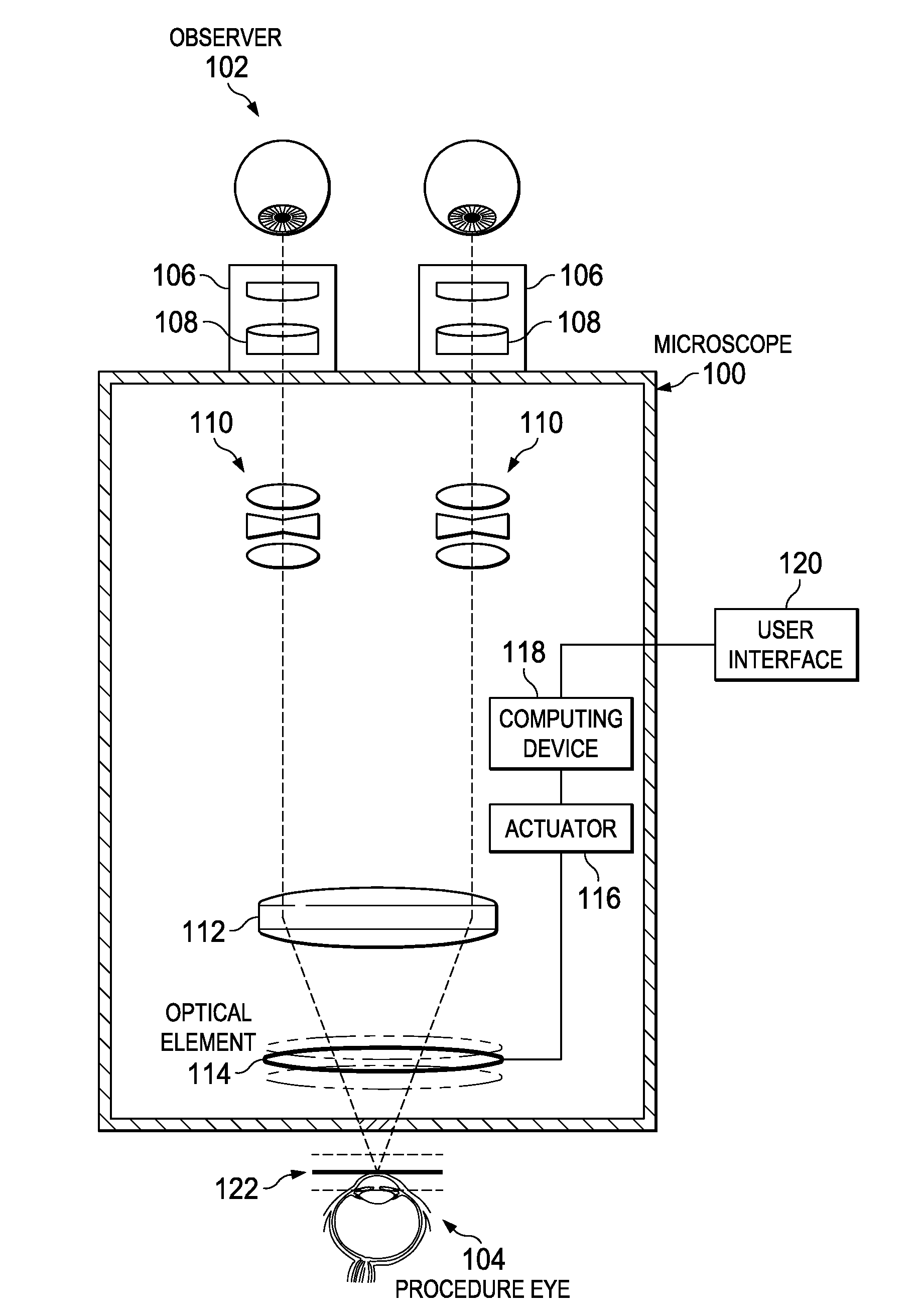
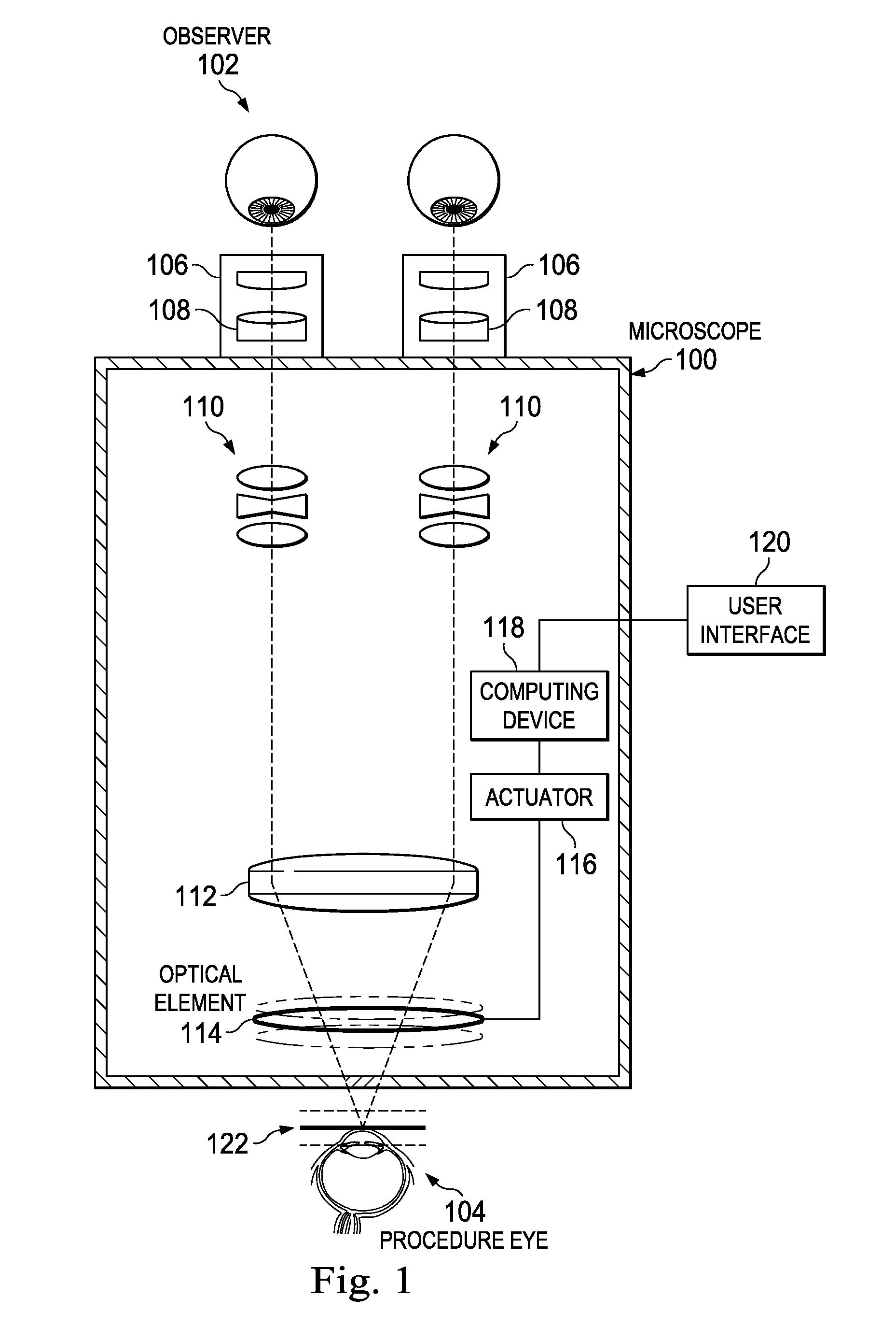
  • Figure 1
    Figure 1
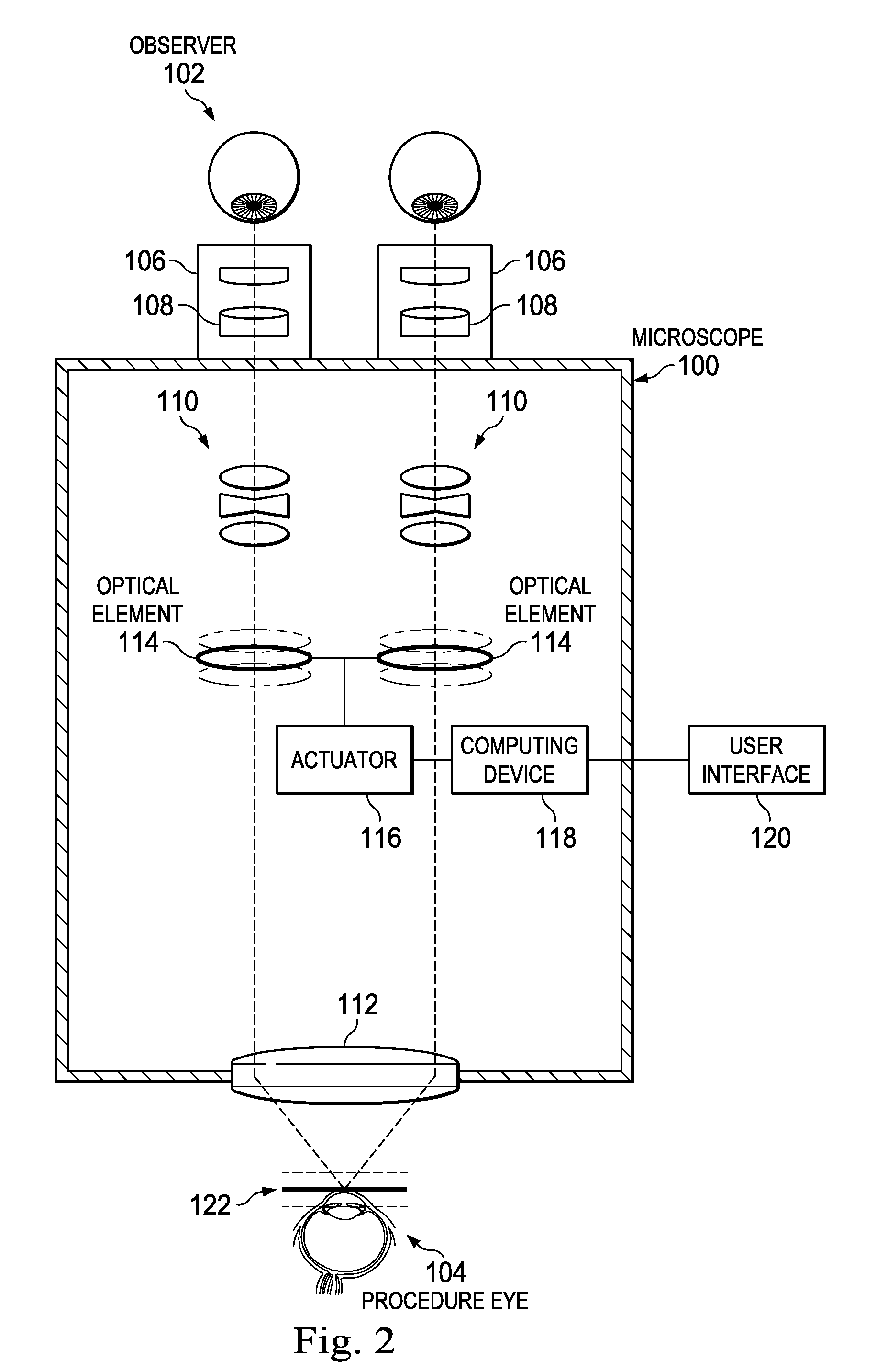
  • Figure 2
    Figure 2
  • Figure 3
    Figure 3
Patent Text Reader

Abstract

An ophthalmic surgical microscope can include a movable optical element positioned in an optical pathway of light reflected from a surgical field. The movable optical element can be configured to oscillate in a direction along the optical pathway. The microscope can include an actuator coupled to the movable optical element and configured to move in response to a control signal. The microscope can include a computing device in communication with the actuator and configured to generate the control signal to move the movable optical element. In some embodiments, the computing device is configured to generate the control signal to move the movable optical element with an oscillation frequency greater than the critical flicker fusion rate.
Need to check novelty before this filing date? Find Prior Art

Description

BACKGROUND

[0001] 1. Technical Field

[0002] Embodiments disclosed herein are related to ophthalmic surgical microscopes. More specifically, embodiments described herein relate to an increased depth of field microscope including a movable optical element.

[0003] 2. Related Art

[0004] Doctors can use surgical microscopes during ophthalmic surgical procedures to see fine details of a patient's eye. A successful procedure can depend on the doctor's ability to view the patient's eye clearly using the microscope. One measure of the doctor's ability to view the patient's eye is the depth of field of the microscope. The depth of field can describe an extent of a microscope subject perceivable by an observer to be in focus at the same time along a direction of the optical pathway of light reflected from the subject. For example, the depth of field can describe the longitudinal extent of a patient's eye that is in focus at the same time for a doctor along the longitudinal axis or z-axis of the micros...

Examples

Embodiment Construction

[0018]In the following description specific details are set forth describing certain embodiments. It will be apparent, however, to one skilled in the art that the disclosed embodiments may be practiced without some or all of these specific details. The specific embodiments presented are meant to be illustrative, but not limiting. One skilled in the art may realize other material that, although not specifically described herein, is within the scope and spirit of this disclosure.

[0019]The present disclosure describes an ophthalmic surgical microscope with a movable optical element. The movable optical element can oscillate along a direction of the optical pathway of light reflected from the surgical field at a frequency greater than the CFF rate. Oscillation of the movable optical element can provide an increased depth of field for the microscope. An observer of the surgical field using the microscope optics can perceive a volumetric image. In some embodiments, an image sensor / camera ...