Phospholipid analogs as diapeutic* agents and methods thereof

a technology of phospholipid analogs and tumors, applied in the field of tumor diagnostic imaging using phospholipid analogs, can solve the problems of limited application of tumor-localizing agents developed so far for oncological applications, inability to afford a conclusive diagnosis, and inability to meet the requirements of invasive and costly biopsy procedures

Active Publication Date: 2013-09-17
CELLECTAR LLC
View PDF35 Cites 26 Cited by
  • Summary
  • Abstract
  • Description
  • Claims
  • Application Information

AI Technical Summary

Problems solved by technology

Unfortunately, conventional imaging techniques such as computerized tomography (CT) and MRI (magnetic resonance imaging) are limited in their ability to afford a conclusive diagnosis of a suspected lesion, since they are only capable of observing differences in the density or morphology of tissues.
A more invasive and costly biopsy procedure is often necessary to provide a definitive diagnosis.
Unfortunately, the tumor-localizing agents developed thus far for oncological applications have had only limited application.
Unfortunately, 67Ga gallium citrate is taken up by a variety of other non-cancerous lesions as well, including inflammatory lesions, and unacceptable amounts of radioactivity can also accumulate in liver and spleen tissue.
The rapid buildup of a radiopharmaceutical in these organs can seriously interfere with the imaging of nearby lesions and also negatively impacts the dosage that can safely be given to a patient.
However, these monoclonal antibodies are specific only to the particular tumor tissue for which they have been produced, and therefore will not localize generally in neoplastic tissue.
Moreover, the use of Mabs for diagnostic imaging has lead to additional problems, including varying degrees of antigen expression, low tumor uptake, non-specific binding and adverse immunogenic reactions.
Unfortunately, the data obtained from these studies has also demonstrated a relatively rapid clearance of the radiopharmaceutical compound from the blood, and an undesirable accumulation by non-target tissues.
As noted above, non-target tissue uptake can decrease the efficacy of radiodiagnostic imaging by creating high background activity, or by causing excessive exposure of radiosensitive tissues to the injected radioactivity.
Further, during treatment and diagnosis using CT scanning and RF ablation, functional information from CT scans is difficult to obtain.
With contrast-enhanced helical CT, the tumor vascularity may be assessed to some degree, but there is no way of accurately determining if viable tumor cells remain within the RF lesion.
This decreases the sensitivity and specificity for early detection of recurrent tumor.
Regardless of its tumor targeting ability, FDG due to its rapid metabolism in tumor cells, does not have potential for therapy.
While some intra-treatment clinical assessments are useful in adjusting treatment,14 in most cases there is no accurate method of predicting tumor response during actual treatment.
These are investigational and none are in routine clinical use.
Despite aggressive treatment with surgery, radiation, and chemotherapy, most patients harboring these tumors have less than a two-year survival after diagnosis.
Most malignant gliomas, however, have an infiltrative component, which is poorly differentiated from edematous brain tissue by present imaging techniques.
It is often this component of the tumor that is most difficult to treat and responsible for local recurrence.
Likewise, pancreatic cancer is a highly lethal disease with the poorest likelihood of survival among all of the major malignancies.
However, it is one of the most highly lethal diseases which accounts for 5% of all cancer deaths.
In addition, although surgical resection offers the only hope for cure, the five-year survival after resection is only 20%.
Although PET scanning with 18-FDG has shown promise in imaging a variety of other primary cancers, it appears to have only limited ability to improve upon the imaging capability of CT scan for patients with pancreatic cancer, particularly in assessing for metastatic disease.
Although resection is the only curative option, only 10-30% of patients are candidates for surgery at the time of presentation, due to either poor hepatic reserve or the presence of unresectable or metastatic disease.
More sophisticated imaging methods including scintigraphic scanning with 99mTc-Sestamibi and 18F-FDG PET scanning have now been extensively examined, but have not impacted treatment planning significantly due mainly to unpredictable specificity.
Additionally, the use of radiation is associated with specific toxicities which are generally late-occurring and dose-limiting.
Fibrosis, nerve damage, and soft tissue necrosis can be severe if excessive doses of radiation are used.
However, this is an expensive imaging test which is not available in most community practices.
Limitations of PET scanning, however, include the cost, limited availability, inability to detect lesions under 1 cm, and lack of specificity, particularly in patients with inflammatory or granulomatous disease.
Conventional anatomic imaging techniques such as CT scanning are also not good at predicting survival following treatment despite tumor shrinkage following therapy.
Although PET imaging procedures are gaining effectiveness in this area, the cost and inaccessibility severely limits its practical application.

Method used


Image

  • Phospholipid analogs as diapeutic* agents and methods thereof
  • Phospholipid analogs as diapeutic* agents and methods thereof
  • Phospholipid analogs as diapeutic* agents and methods thereof

Examples


example i

A. Example I

Synthesis, Radiolabeling, and Formulation of NM404

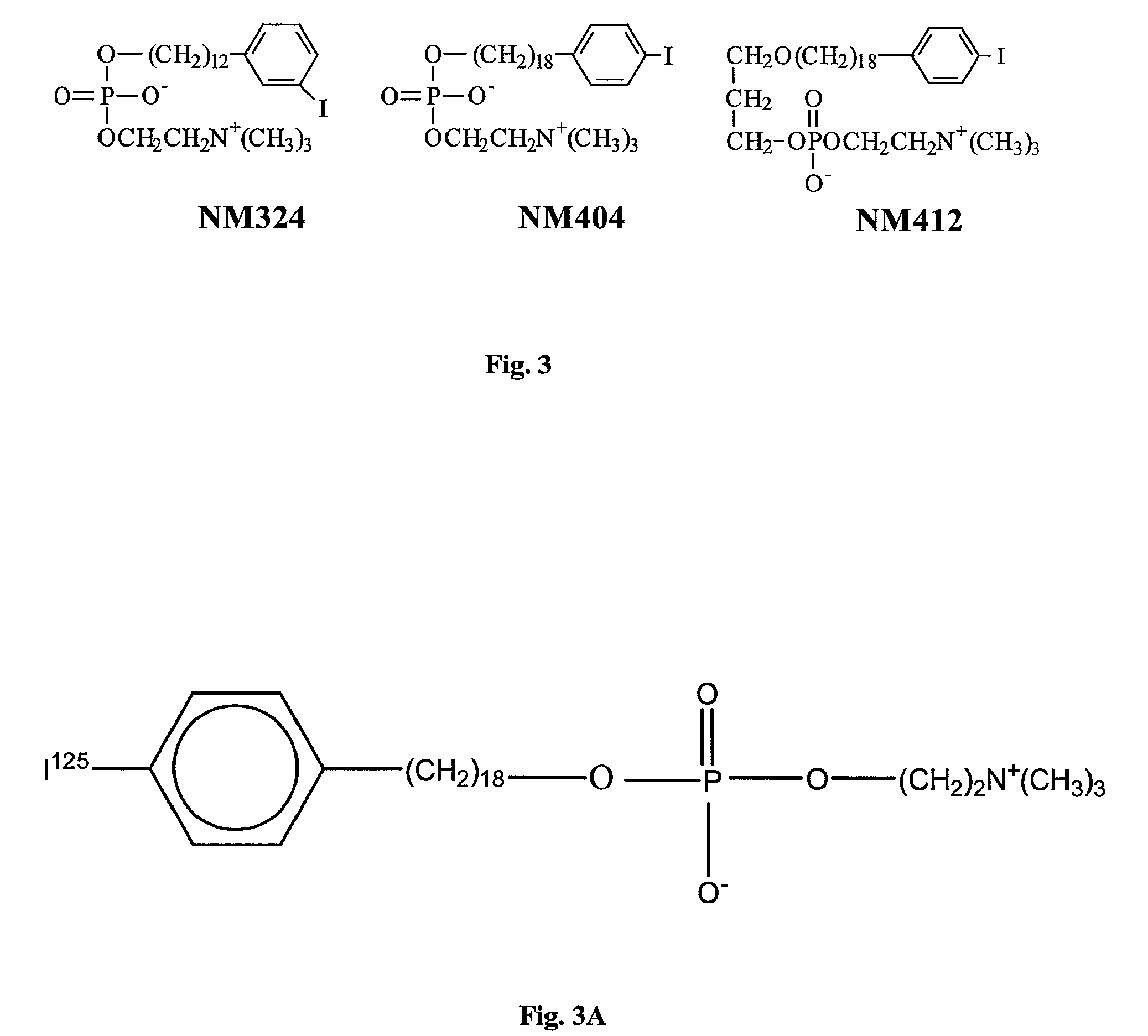
[0126]The inventors' synthetic approach was based on the copper-catalyzed cross-coupling reaction of Grignard reagents with alkyl tosylates or halides for the alkyl chain elongation (see the scheme below). The synthesis was started from p-iodobenzyl alcohol 1 which was converted into p-iodobenzyl bromide 2 by reaction with trimethylsilyl bromide. p-lodobenzyl bromide 2 was further coupled with Grignard reagent 3 in the presence of Li2CuCl4 as a catalyst. 12-(p-lodophenyl)dodecanol 5 obtained after deprotection of the first coupling product 4 was converted into tosylate 6. In the next step, tosylate 6 was coupled with Grignard reagent 7 containing 6 carbon atoms and this completed the chain elongation process. THP deprotection of 8 gave 18-(p-iodophenyl)octadecanol 9 which was converted into 10 (NM-404) by two-step procedure as shown in the scheme.

[0127]

[0128]Further, rapid high yield synthesis process for labeling NM404 w...

example ii

B. Example II

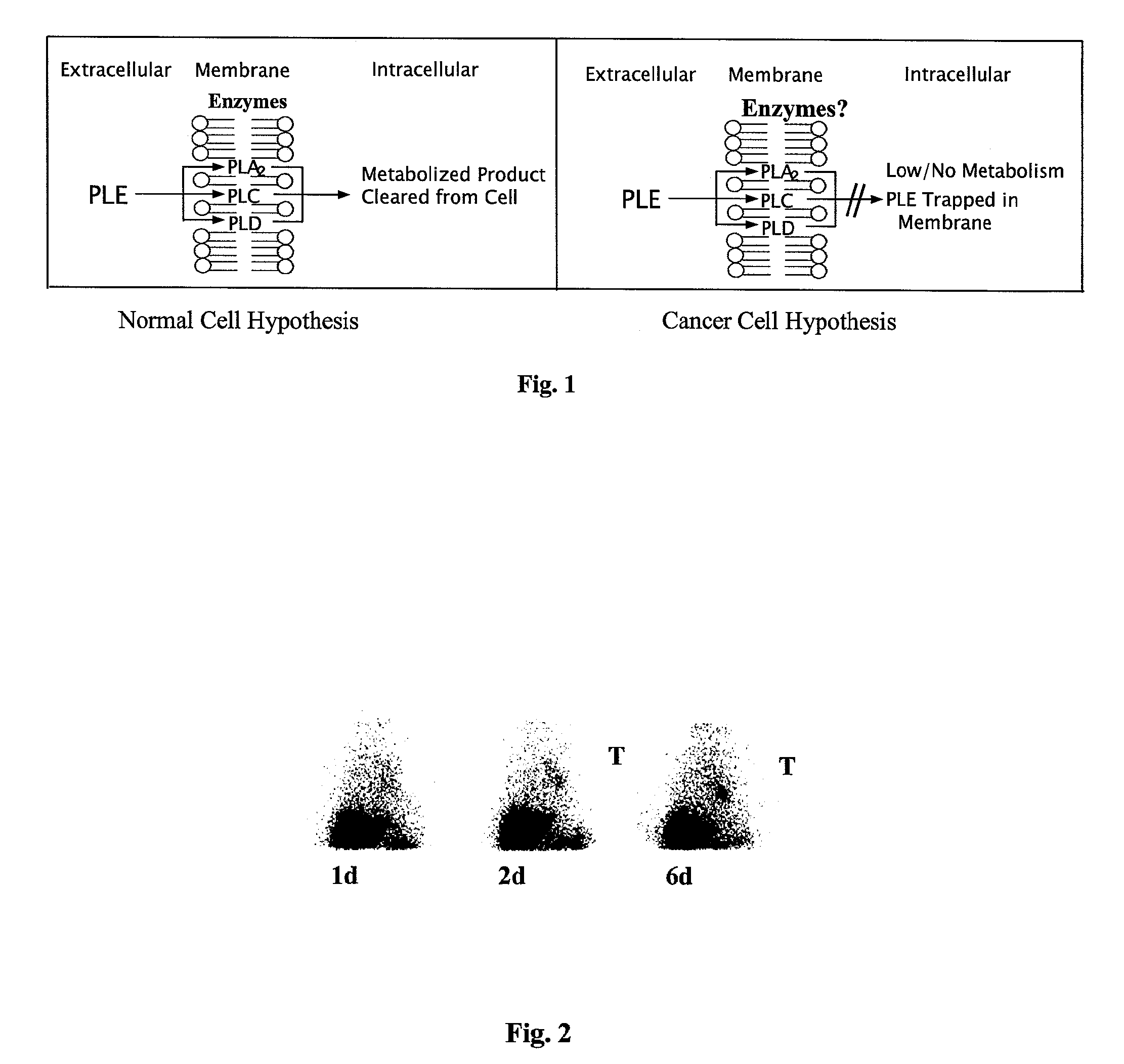
Preclinical Studies with First Generation PLE Analogs

[0144]Phospholipid ethers can easily be labeled with iodine radioisotopes using an isotope exchange method developed by the inventors.19 The iodophenyl phospholipid ether analogs are specifically designed so that the radioiodine affixed to each molecule is stable to facile in vivo deiodination. Over 20 radiolabeled PLE compounds were synthesized and tested in vitro and in vivo.20-22 Two of these, namely NM294 and NM324 [12-(3-iodophenyl)-dodecyl-phosphocholine], initially showed the most promise in animal tumor localization studies. These prototype compounds, labeled with iodine-125, selectively localized in tumors over time in the following animal tumor models; 1) Sprague-Dawley rat bearing Walker 256 carcinosarcoma; 2) Lewis rat bearing mammary tumor; 3) Copenhagen rat bearing Dunning R3327 prostate tumors; 4) Rabbits bearing Vx2 tumors; and 5) athymic mice bearing human breast (HT39), small cell lung (NCl-69), colo...

example iii

C. Example III

Non-Small Cell Lung Cancer

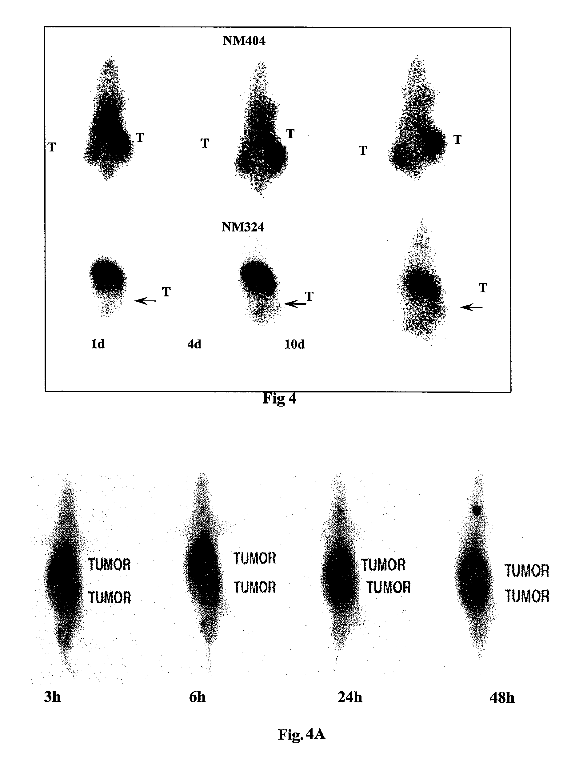
[0157]Imaging and biodistribution studies were performed in SCID (severe combined immune deficiency mutation) mice bearing the human NSCLC adenocarcinoma A549 cell line (adenocarcinoma has become the most frequent human lung cancer histologic type). Preliminary pilot results in five animals were encouraging and demonstrated that the new agent NM404 overcomes a limitation of the NM-324 compound. While there is reasonably good tumor uptake with NM324, imaging is compromised by a high first pass clearance by the liver. However, NM404 shows excellent tumor visualization, especially striking in the delayed images, with minimal liver and kidney uptake. Moreover, tissue biodistribution studies further confirmed the high levels of radioactivity residing in the tumors. A comparison of NM324 and NM404 images in a SCID mouse-human NSCLC model are shown in FIG. 4. Note the relative lack of liver, kidney and gut activity with NM404, coupled with excellent ...


PUM


Abstract

The present invention provides methods and uses of phospholipid ether analogs as diagnostic and therapeutic agents for numerous cancers.

Description

RELATED APPLICATION[0001]The present application seeks priority from U.S. Provisional Application 60 / 521,166, filed on Mar. 2, 2004, which is incorporated herein by reference for all purposes.BACKGROUND OF INVENTION[0002]The invention generally relates to diagnostic imaging of tumors and specifically relates to diagnostic imaging of tumors using phospholipid analogs.[0003]The early detection of cancer has been one of the primary goals of modern imaging technology, since the identification of a suspected tumor in a localized stage significantly improves the chances for successful treatment and elimination of the cancerous tissue. A large number of imaging strategies have therefore been designed, using a variety of techniques and modalities, to aid the physician in making an accurate diagnosis as early as possible.[0004]Unfortunately, conventional imaging techniques such as computerized tomography (CT) and MRI (magnetic resonance imaging) are limited in their ability to afford a concl...

Claims


Application Information

Patent Timeline
Patent Type & Authority Patents(United States)
IPC IPC(8): A61K51/00A61M36/14A61K49/00A61K51/04
CPCA61K51/04C12Q1/34A61K51/0408G01N33/57492C07F13/005A61K49/0017A61P35/00A61P43/00A61K51/0489
Inventor WEICHERT, JAMEYLONGINO, MARCPINCHUK, ANATOLY
Owner CELLECTAR LLC
Why Eureka
Try Eureka