
Scaffold-free self-organized 3D synthetic tissue
a self-organized, 3d technology, applied in the field of regenerative medicine, can solve the problems of insufficient prediction of the influence of organism safety, fragile single sheet obtained by this technique, and insufficient provision of extracellular matrix, so as to achieve reliable and safe cell implantation
Patent Information
- Authority / Receiving Office
- US · United States
- Patent Type
- Applications(United States)
- Current Assignee / Owner
- Publication Date
- 2008-01-03
Smart Images
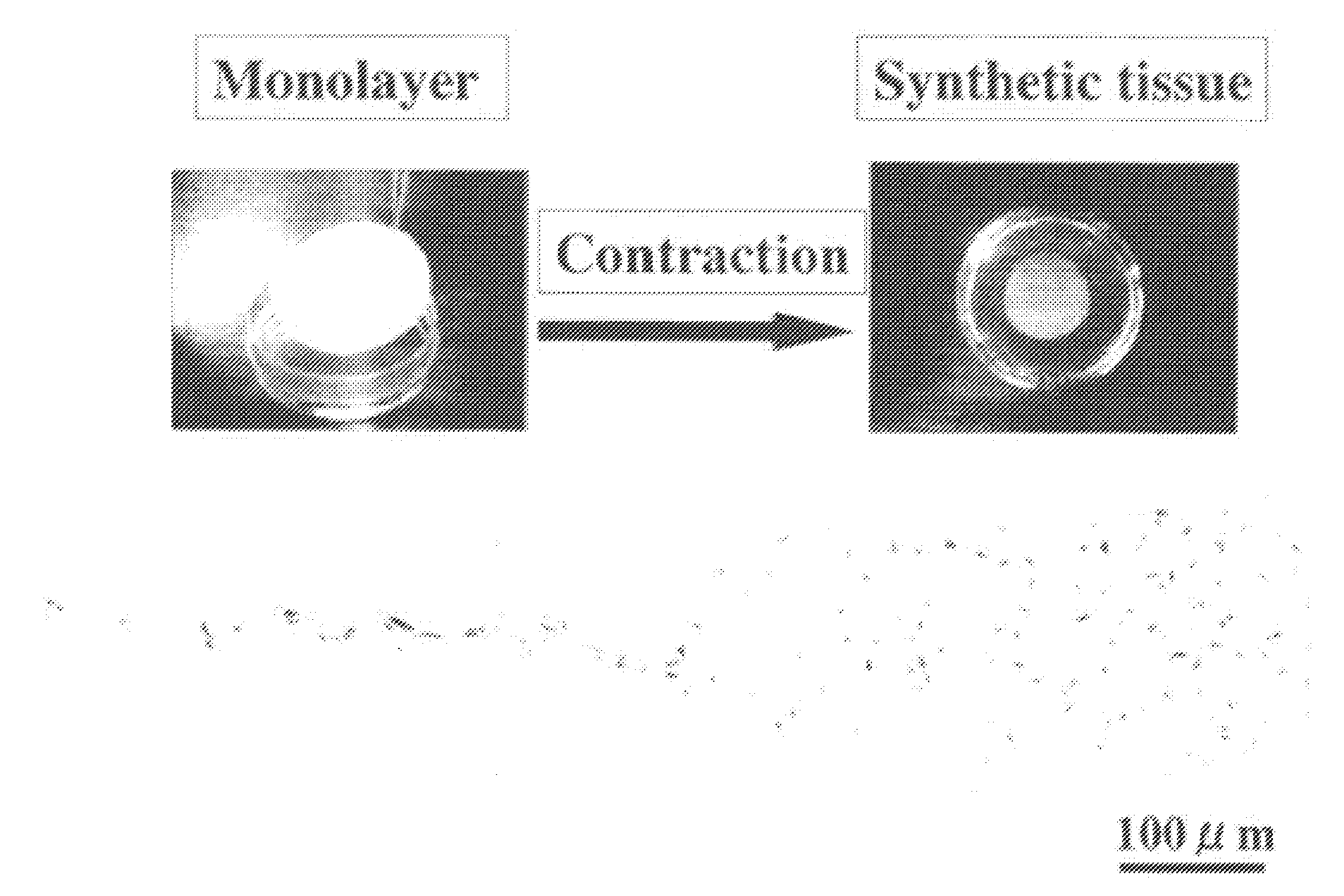
Figure 1 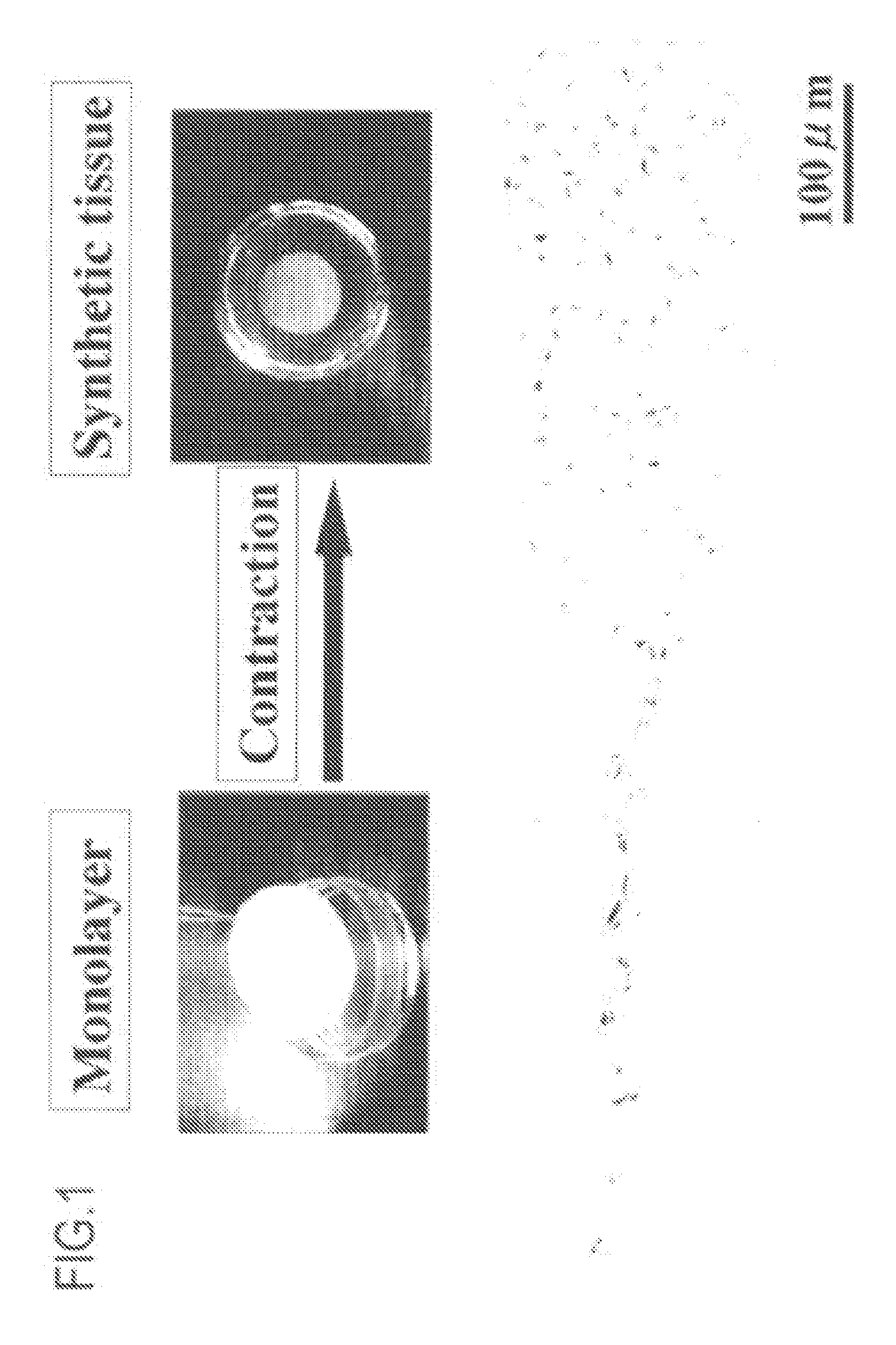
Figure 2 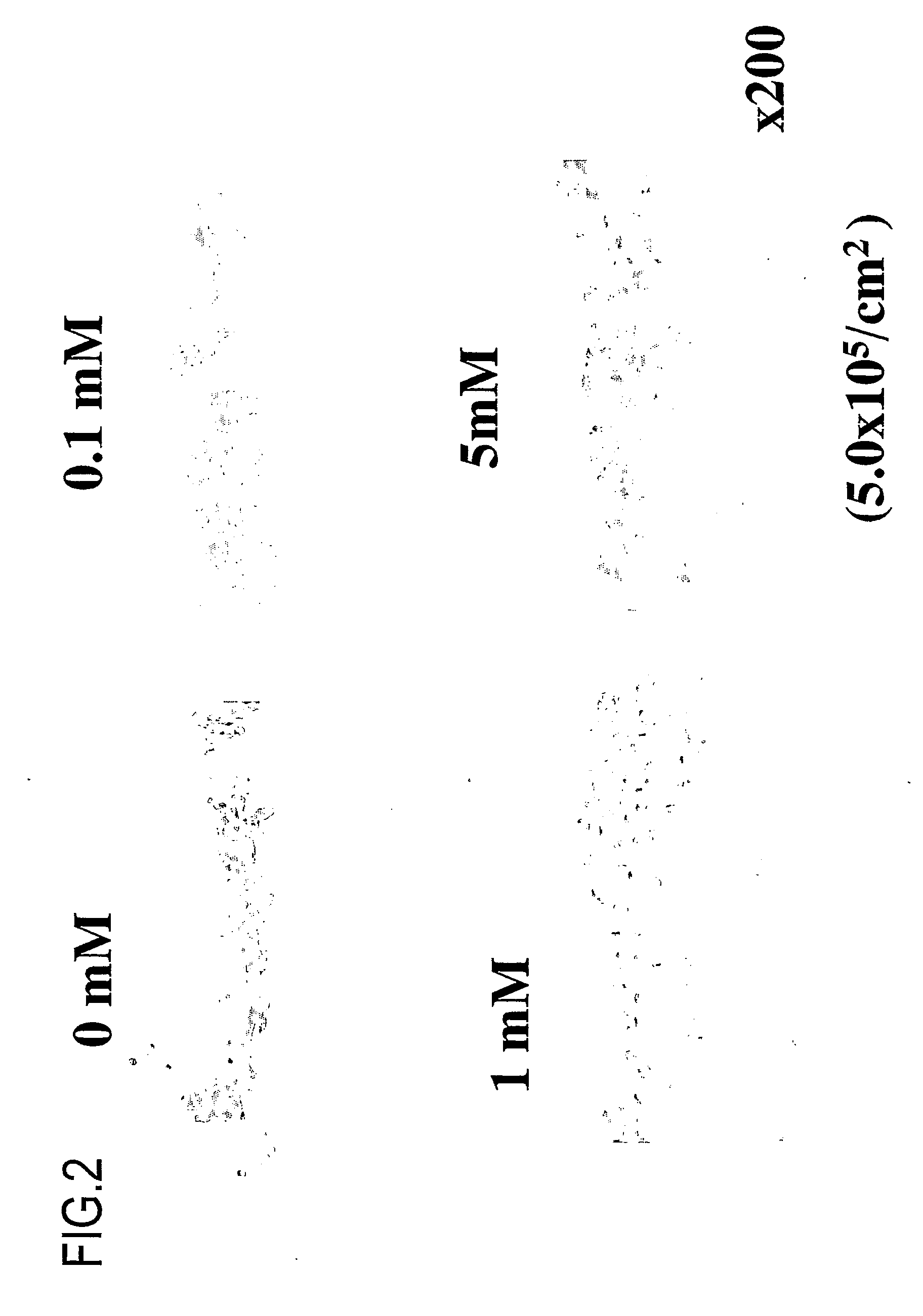
Figure 3
Abstract
Description
CROSS-REFERENCE TO RELATED APPLICATION
[0001] This application is a continuation of U.S. patent application Ser. No. 10 / 566,845, now pending, which application is a U.S. National stage application of PCT / JP2004 / 011401, international filing date of Aug. 2, 2004, which applications are incorporated herein by reference in their entireties.STATEMENT REGARDING SEQUENCE LISTING
[0002] The Sequence Listing associated with this application is provided in text format in lieu of a paper copy, and is hereby incorporated by reference into the specification. The name of the text file containing the Sequence Listing is 690128—401C1_SEQUENCE_LISTING.txt. The text file is 313 KB, was created on Feb. 16, 2007, and is being submitted electronically via EFS-Web, concurrent with the filing of the specification. TECHNICAL FIELD
[0003] The present invention relates to the field of regenerative medicine. More particularly, the present invention relates to a synthetic tissue capable of functioning after im...
Examples
example 1
[0423] In this example, various synovial cells were used to produce a synthetic tissue as follows.
[0424]
[0425] Synovial cells were collected from a knee joint of a pig (LWD ternary hybrid, 2-3 months old upon removal of cells), followed by treatment with collagenase. The cells were cultured and subcultured in 10% FBS-DMEM medium (FBS was obtained from HyClone, DMEM was obtained from GIBCO). It has been reported that 10th passage synovial cells still have pluripotency. Although cells of 10 or less passages were used in this example, cells of more than 10 passages may be used depending on the application. Autotransplantation was performed for humans, where a sufficient number of cells were used and the cells were cultured for a short period of time so as to reduce the risk of infection or the like.
[0426] Considering these points, cells of various passages were used. Actually, primary culture cells, first passage cells, second passage cells, third passage cells, fourth ...
example 2
Measurement of Collagen Production
[0444] Next, it was determined whether or not collagen (extracellular matrix) is sufficiently secreted after implantation of a synthetic tissue of the present invention.
[0445] The following protocol was used.
[0446]
[0447] Culture periods: 3 days, 7 days, 14 days, and 21 days,
[0448] Concentrations of ascorbic acid 2-phosphate: 0 mM, 0.1 mM, 1 mM, and 5 mM
[0449] Under the above-described conditions, a synovial membrane-derived synthetic tissue was produced.
[0450] 6 N HCl was added to culture medium for the synthetic tissue, followed by hydrolysis at 105° C. for 18 hours. The medium was oxidized with chloramine T. Thereafter, the synthetic tissue was subjected to color development using Ehrlich's Reagent Solution (2 g of p-dimethylamino-benzaldehyde+3 ml of 60% perchloric acid; isopropanol was diluted at 3:13), followed by measurement of absorbance.
[0451]
[0452] 1) The quantities of collagen produced was dependent on the ascorbic acid concentratio...
example 3
Influences of the Size of a Dish, the Number of Cells, and the Number of Passages
[0454] Next, influences of the size of a dish and the number of passages were investigated.
[0455]FIG. 9 shows the formation of synthetic tissues where the number of cells and the number of the passage were changed. A synthetic tissue was formed in all concentrations tested.
[0456] Under the conditions of the above-described Example 1, a similar experiment was conducted where the sizes of dishes were 35 mm, 65 mm, and 100 mm and the number of passages were 5 to 7 (FIG. 10).
[0457] The results are shown in FIGS. 9 and 10. FIG. 9 shows the states of synthetic tissues, where the number of passages was changed. FIG. 10 shows the states of synthetic tissues, where the size of a dish was changed. As can be seen from the figures, it was demonstrated that a synthetic tissue can be formed using any size of dish and any number of passages.
[0458] As shown in FIG. 9, basically, a greater number of cells may be pr...