
Method for assaying the activity of lysosomal enzymes
a technology of lysosomal enzymes and assay methods, which is applied in the field of assaying the activity of lysosomal enzymes, can solve the problems of not having the therapy available for brain disorders, requiring risky and costly therapeutic procedures, and progressing irreparable damage to the brain
Patent Information
- Authority / Receiving Office
- US · United States
- Patent Type
- Applications(United States)
- Current Assignee / Owner
- Publication Date
- 2009-12-31
- Estimated Expiration
- Not applicable · inactive patent
Smart Images
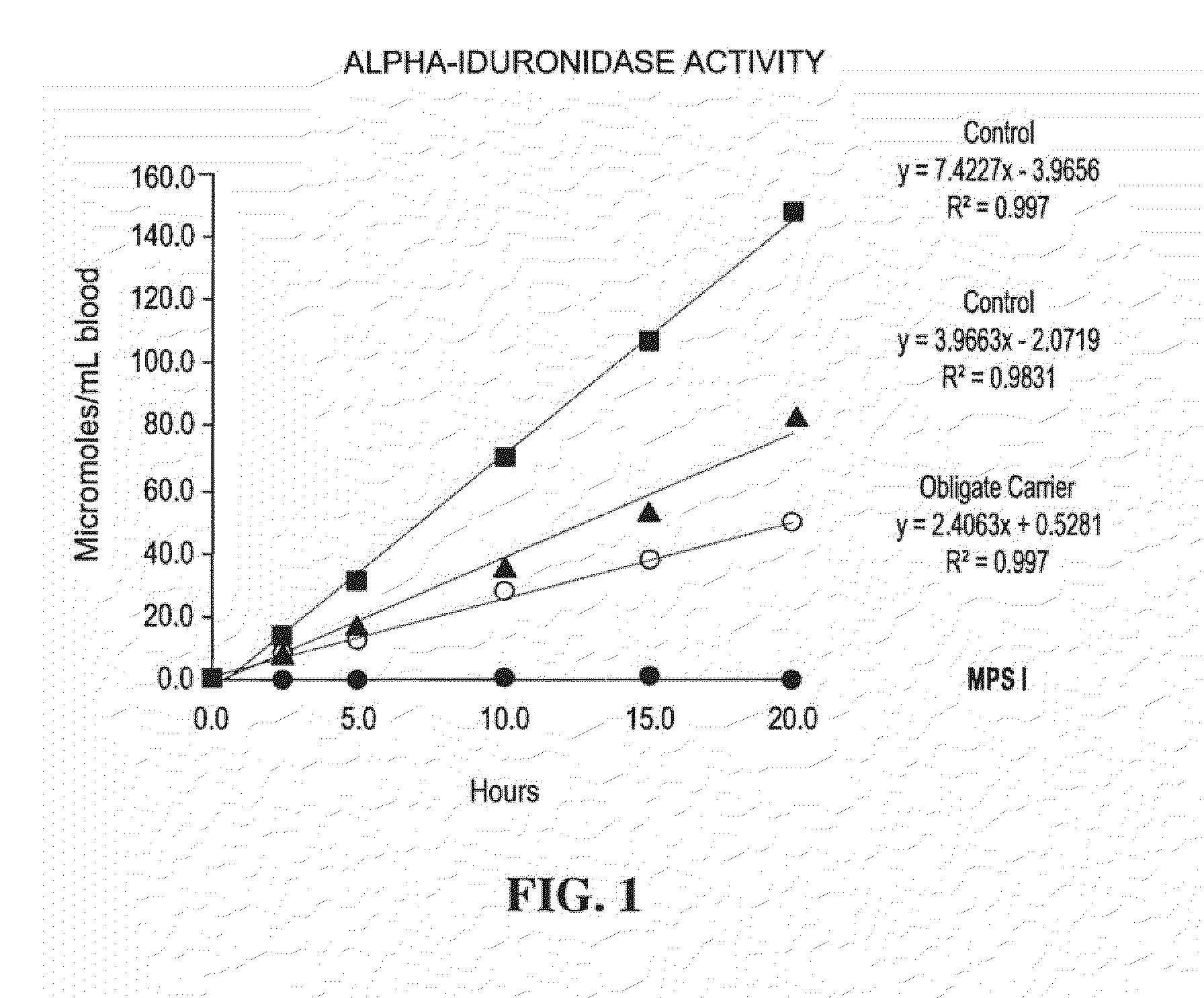
Figure 1 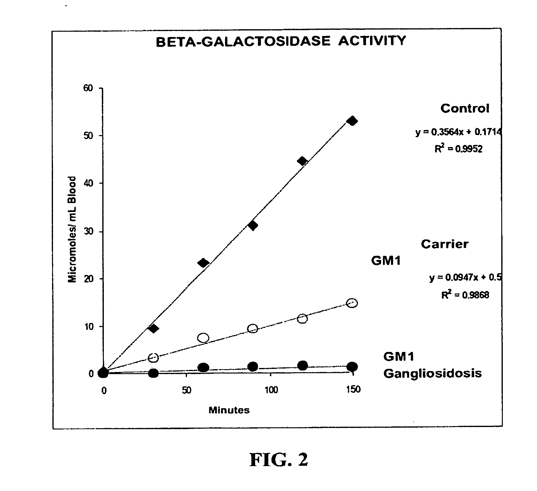
Figure 2 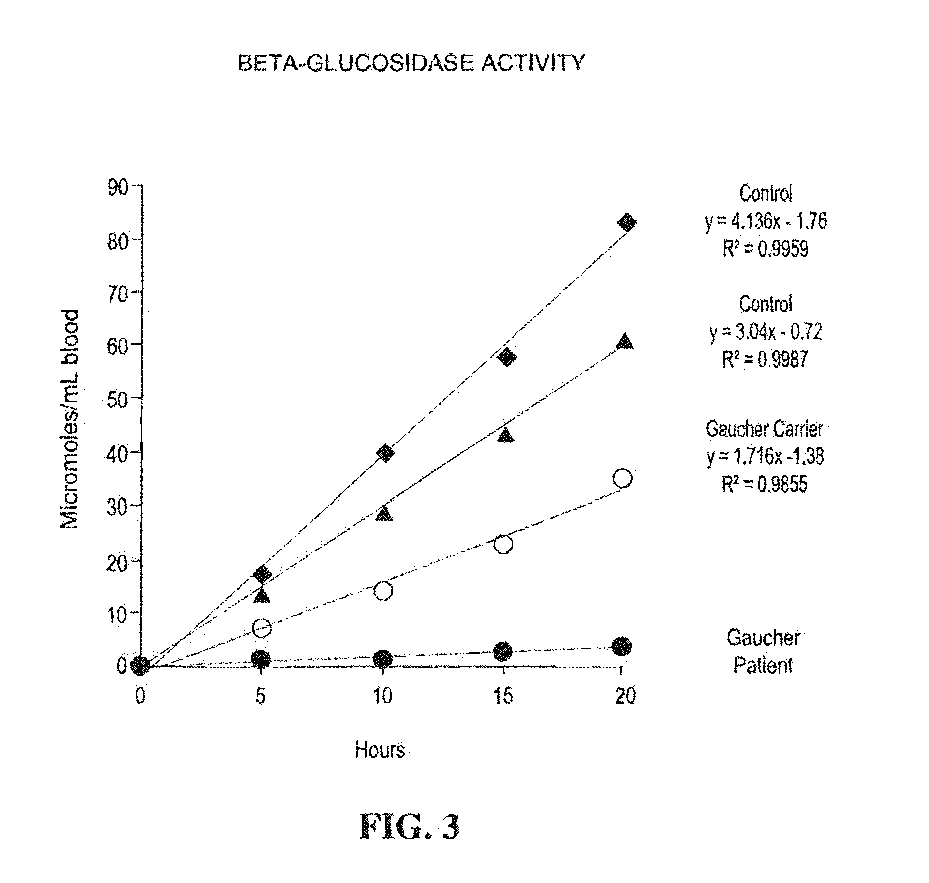
Figure 3
Abstract
Description
[0001] This application claims the benefit of U.S. Provisional Application No. 60 / 227,573, filed Aug. 25, 2000, and Patent Cooperation Treaty Application No. PCT / US01 / 26259 filed Aug. 24, 2001, which are incorporated entirely by reference.TECHNICAL FIELD OF THE INVENTION
[0002] This invention relates to a method of assaying lysosomal enzymes present in dried samples of bodily fluids and cell tissues. The invention also relates to a diagnostic kit for assaying lysosomal enzymes present in dried samples of bodily fluids and cell tissues.BACKGROUND OF THE INVENTION
[0003] The lysosome is an organelle founded in the cytoplasm of eukaryotic cells, which serves as storage for many hydrolytic enzymes and as a center for degrading and recycling cellular components. This organelle contains several types of hydrolytic enzymes, including proteases, nucleases, glycosidases, lipases, phospholipases, phosphatases and sulfatases. All enzymes are acid hydrolases. See Lehninger et als. Principles of Bioc...
Examples
example 1
Preparation of Dried Blood Samples
[0099]A drop of blood obtained by venepuncture was spotted on filter paper (Schleicher and Schuell No 903, Keene, N.H., USA) and allowed to dry at room temperature (22° C.) overnight on a flat non-absorbing surface. The dried blood spots on filter paper were stored on plastic bags at 4° C. until analysis.
[0100]A sample for analysis was prepared by punching out a 3 mm-diameter circle (about 5.5 μl of whole blood) with a standard paper punch from the dried blood spots on the filter paper. A sample for analysis was prepared by punching out a 1.5 mm-diameter circle (about 2 μl of whole blood) with a standard paper punch from the dried blood spots on the filter paper.
[0101]This protocol was also performed by utilizing a heelprick finger to obtain the blood.
example 2
Preparation of Dried Chorionic Villae Samples
[0102]A chorionic villae sample (about 10 mg) was sonicated twice by 20 seconds in 50 μl of cold distilled water (Heat Systems-Ultrasonics, Inc., model W225R).
[0103]After removing 10 μl for protein determination, the sonicated cells were spotted on filter paper (Schleicher and Schuell No 903, Keene, N.H., USA) and allowed to dry for 6 hours at room temperature (22° C.) on a flat non-absorbing surface. See Lowry et al., J. Biol. Chem. 193:265-275 (1951). The spotted filter paper was stored on plastic bags at −20° C. until analysis.
[0104]A sample for analysis was prepared by punching out a 3 mm-diameter circle with a standard paper punch from the dried chorionic villae spots on the filter paper.
example 3
Preparation of Dried Cultured Amniocytes Samples
[0105]Cultured amniocytes were suspended in 500 μl of cold phosphate saline buffer (pH 7.4). After centrifugation at 1,200 g for 5 minutes at 4° C. the supernatant was removed by aspiration. The cell pellet was resuspended in 40 μl of cold distilled water and sonicated (Heat Systems-Ultrasonics, Inc., model W225R).
[0106]After removing 10 μl for protein determination, the sonicated cells were spotted on filter paper (Schleicher and Schuell No 903, Keene, N.H., USA) and allowed to dry for 6 hours at room temperature (22° C.) on a flat non-absorbing surface. See Lowry et al., supra. The spotted filter paper was stored on plastic bags at −20° C. until analysis.
[0107]A sample for analysis was prepared by punching out a 3 mm-diameter circle with a standard paper punch from the dried amniocytes spots on the filter paper.