
Ultra-Low Damping Imaging Mode Related to Scanning Probe Microscopy in Liquid
a scanning probe and liquid imaging technology, applied in the field of new imaging mode for scanning probe microscopy, can solve the problems of reducing the q factor of the afm probe, unable to extract meaningful data, and affecting image resolution, so as to achieve high resolution, good resolution, and low damping
Patent Information
- Authority / Receiving Office
- US · United States
- Patent Type
- Applications(United States)
- Current Assignee / Owner
- Publication Date
- 2012-11-01
- Estimated Expiration
- Not applicable · inactive patent
Smart Images
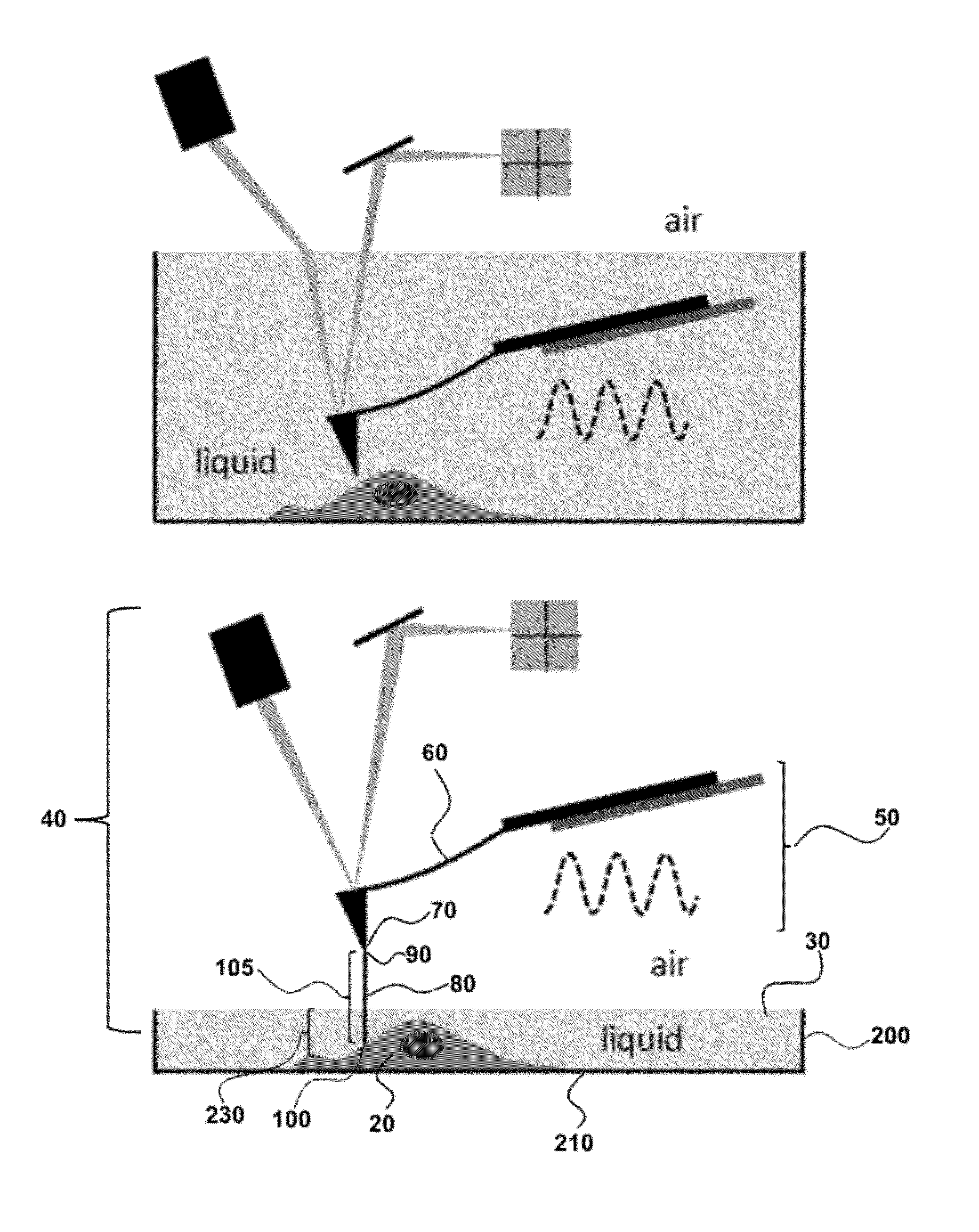
Figure 1 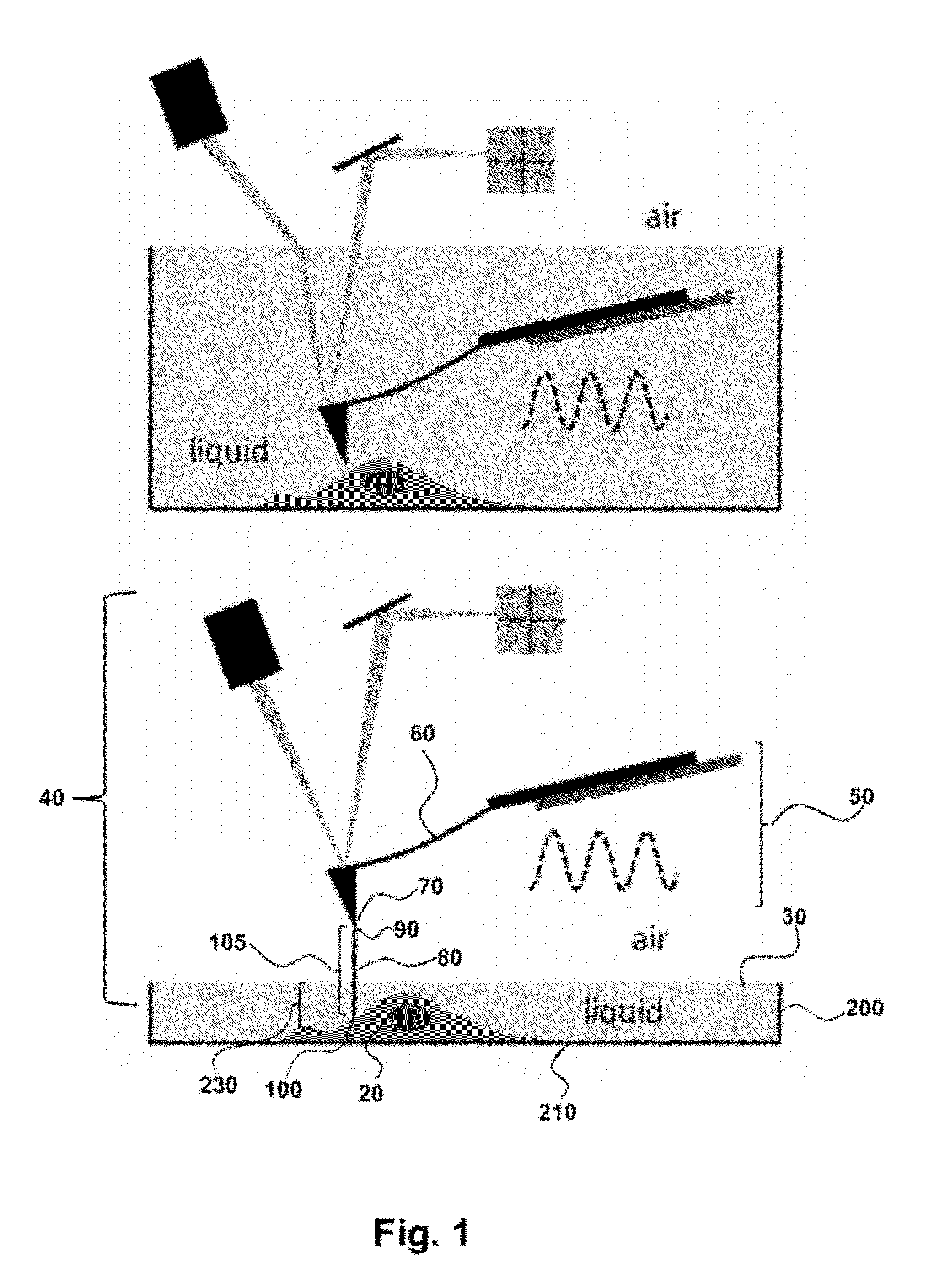
Figure 2 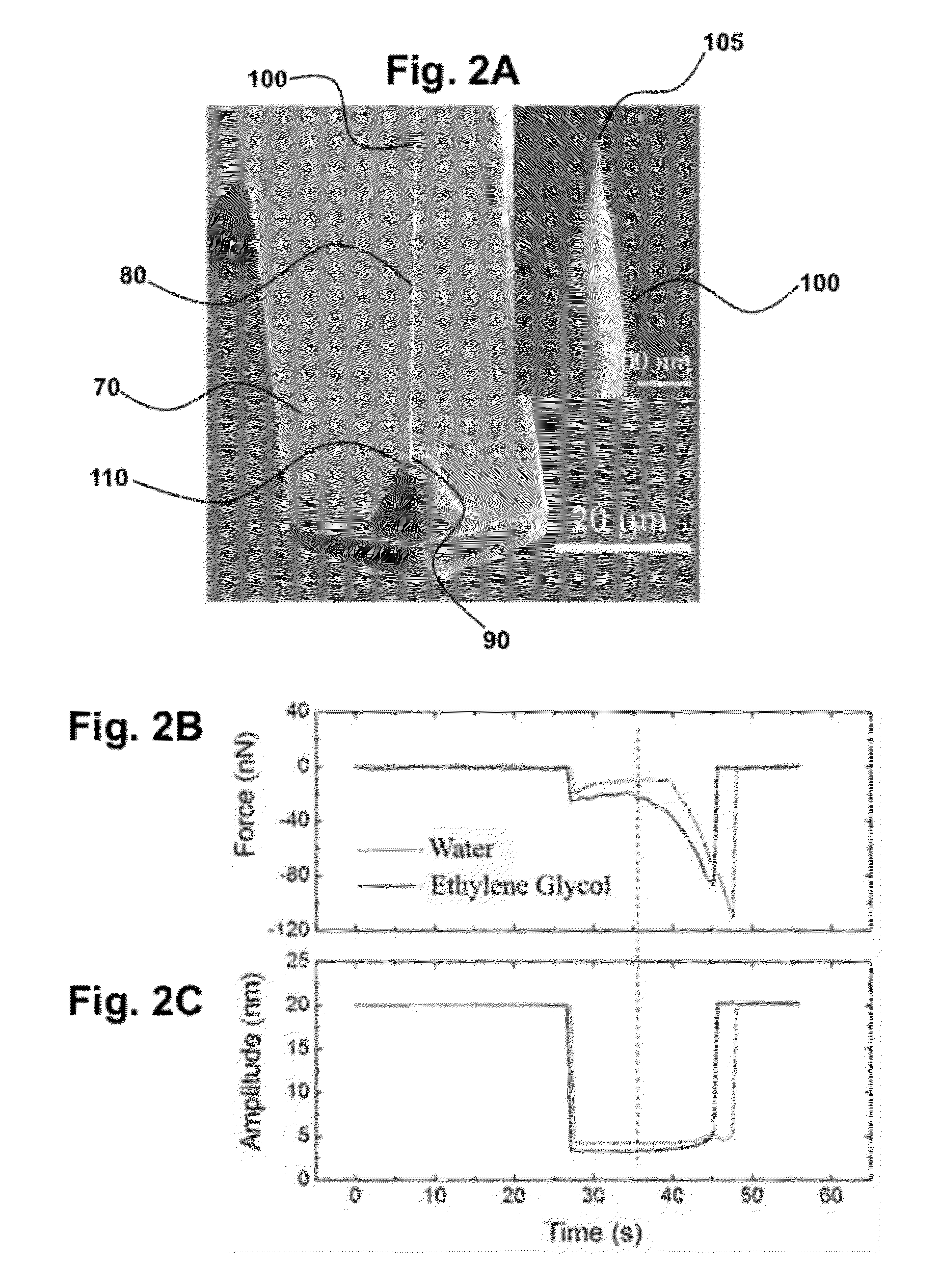
Figure 3
Abstract
Description
STATEMENT REGARDING FEDERALLY SPONSORED RESEARCH OR DEVELOPMENT
[0001] This invention was made with government support under CBET 0731096 awarded by the National Science Foundation. The government has certain rights in the invention.CROSS-REFERENCE TO RELATED APPLICATIONS
[0002] Not applicable.BACKGROUND OF INVENTION
[0003] Methods and devices provided herein relate to a new imaging mode for scanning probe microscopy in a liquid environment. The specific design and methodology, for example, provides atomic force microscopy (AFM) imaging of a material covered in liquid, where the AFM probe, when dynamically operated, experiences a significantly lowered damping and has a corresponding increase in Q factor when compared to conventional systems, thereby achieving high sensitivity and high resolution dynamic imaging of samples in liquid.
[0004] Conventional AFM systems, when imaging material in liquid, submerse the cantilever portion of the AFM probe in the liquid. The relatively large footprint...
Examples
example 1
[0063]Intrinsically High Q-Factor “Trolling Mode” Scanning Probe Microscopy in Liquid; System Characterization. To be able to interact and image liquid-like cell membranes with high spatial and force resolutions is of significant value for biological studies. The widely used dynamic mode atomic force microscopy (AFM) suffers severe sensitivity degradation and noise increase when operated in a liquid medium. We introduce an alternative imaging scheme with the use of an AFM nanoneedle probe that lowers both the hydrodynamic damping and the thermal fluctuation force experienced by a typical AFM probe by orders of magnitude and acquires an intrinsic Q-factor over 100 without Q-control feedback when operated for AFM imaging in liquid. This allows truly gentle imaging of demanding samples such as the soft membranes of living cells under physiological conditions even with a rigid AFM probe having a force constant over several Newton per meter and a resonance frequency over 200 kHz, and dir...
example 2
[0076]Imaging Materials. The “trolling mode” AFM microscopy is implemented for imaging biological samples in liquid. We prepare the biological samples (collagen fibrils and living HeLa cells) in concave glass containers as mentioned above. We first advance the nanoneedle towards the liquid surface near the wet perimeter while monitoring the amplitude and deflection of the AFM cantilever continuously. The reach of the liquid surface is identified by observing the snap-in motion of the cantilever in which the oscillation amplitude of the cantilever dropped noticeably. We then perform a new tuning procedure with the nanoneedle partially immersed in liquid to precisely determine the new resonance frequency of the nanoneedle cantilever system. To engage onto the sample surface immersed underneath a shallow liquid layer, we rely on identifying the transition in the change of the oscillation amplitude versus the distance dependence. The change in oscillation amplitude is more abrupt when t...