
Microscope, method of operating a microscope and method of imaging a sample
Patent Information
- Authority / Receiving Office
- US · United States
- Current Assignee / Owner
- Publication Date
- 2020-07-23
Smart Images
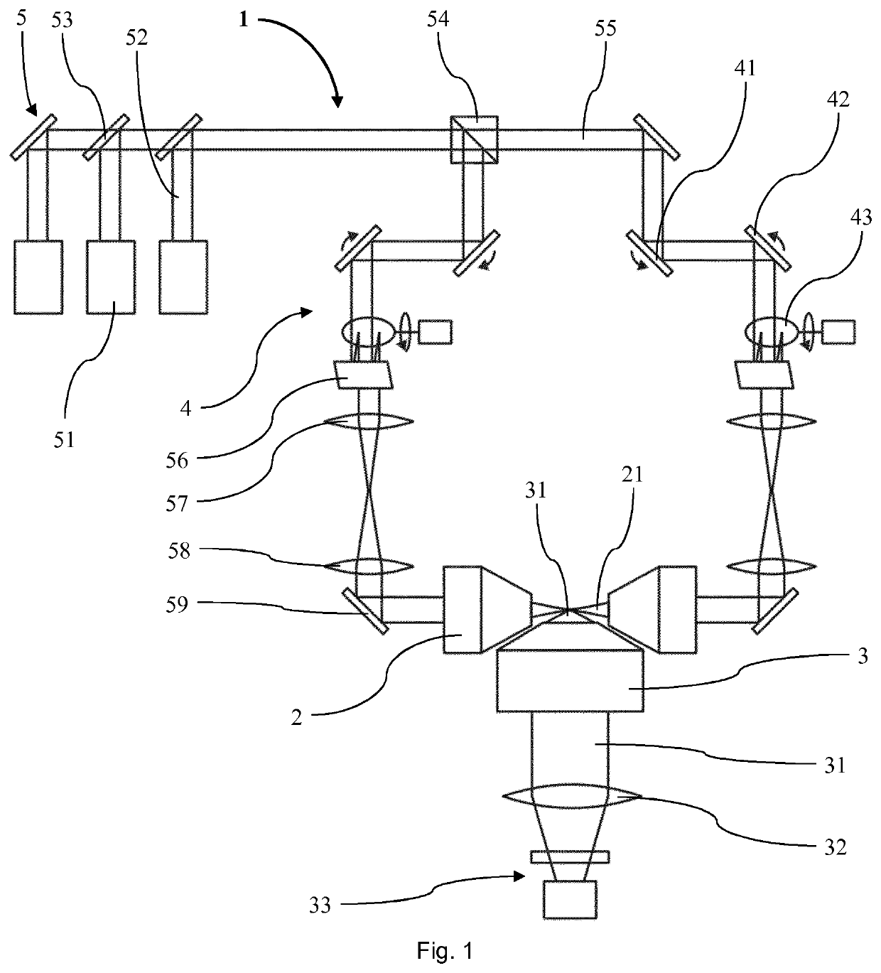
Figure 1 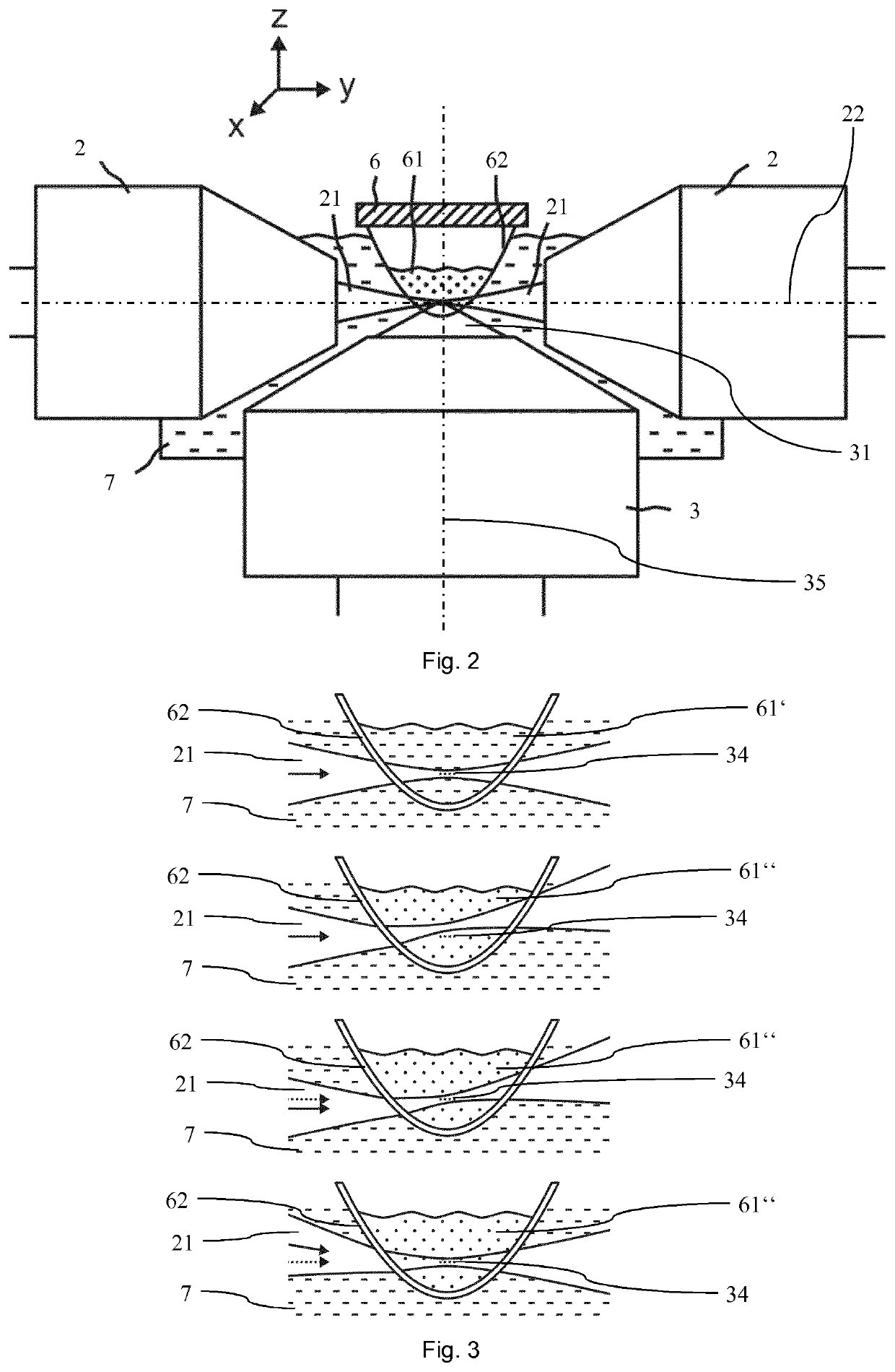
Figure 2
Abstract
Description
TECHNICAL FIELD
[0001] The present invention relates to a microscope according to the preamble of independent claim 1 and more particularly to a method of operating a microscope, a method of imaging a sample and a computer program for operating a microscope. Such a microscope with at least one illumination objective arranged to eject an illumination light beam along an illumination path to illuminate a sample, and an imaging objective arranged to receive detection light comprising at least a portion of the light ejected from the sample, can be used for imaging and analysing the sample.BACKGROUND ART
[0002] Light Sheet (LS) or Selective Plane Illumination Microscopy (SPIM) is a fluorescence microscopy method in which an illumination beam path (excitation light) and a detection beam path (emission light from the sample) are substantially perpendicular to each other. The sample is placed at an intersection of these paths in a focal plane of the imaging objective. For optimal image quality ...
Examples
Embodiment Construction
[0050]In the following description certain terms are used for reasons of convenience and are not intended to limit the invention. The terms “right”, “left”, “up”, “down”, “under” and “above” refer to directions in the figures. The terminology comprises the explicitly mentioned terms as well as their derivations and terms with a similar meaning. Also, spatially relative terms, such as “beneath”, “below”, “lower”, “above”, “upper”, “proximal”, “distal”, and the like, may be used to describe one element's or feature's relationship to another element or feature as illustrated in the figures. These spatially relative terms are intended to encompass different positions and orientations of the devices in use or operation in addition to the position and orientation shown in the figures. For example, if a device in the figures is turned over, elements described as “below” or “beneath” other elements or features would then be “above” or “over” the other elements or features. Thus, the exempla...