
Method of producing cardiac tomogram and tomograph using x-ray ct apparatus
a tomograph and x-ray technology, applied in tomography, applications, instruments, etc., can solve the problems of difficult to highly raise the scan speed to a speed of all heartbeat cycles or less, short projection data of the desired slicing position, and undesirable images. , to achieve the effect of fewer motion artifacts
Patent Information
- Authority / Receiving Office
- US · United States
- Patent Type
- Patents(United States)
- Current Assignee / Owner
- Publication Date
- 2006-02-28
- Estimated Expiration
- Not applicable · inactive patent
Smart Images
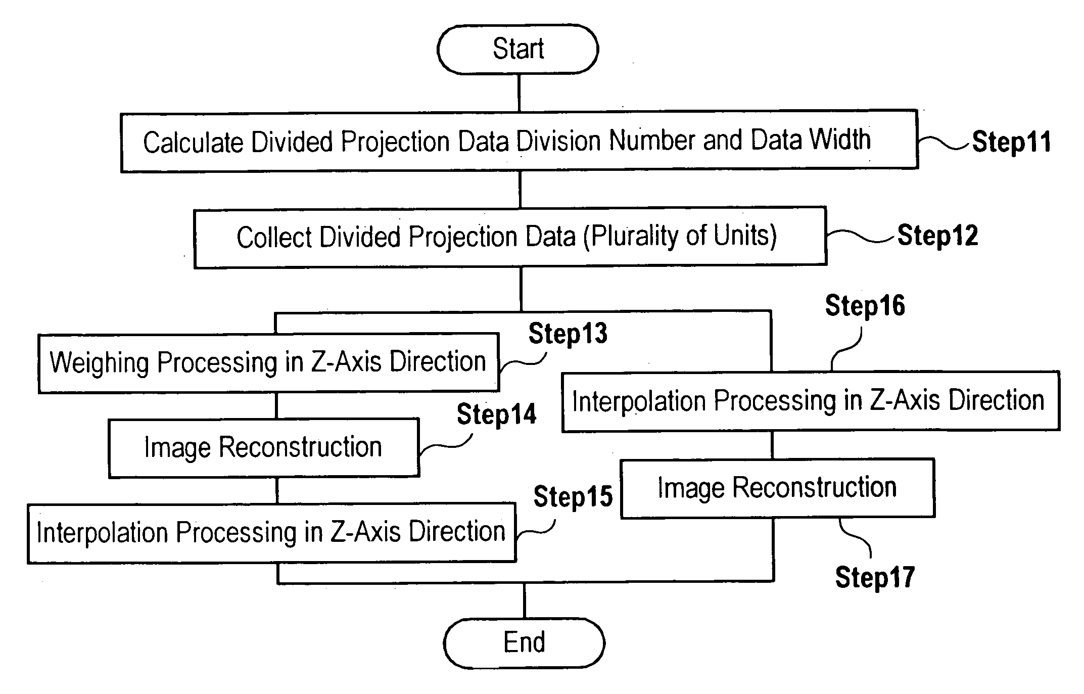
Figure 1 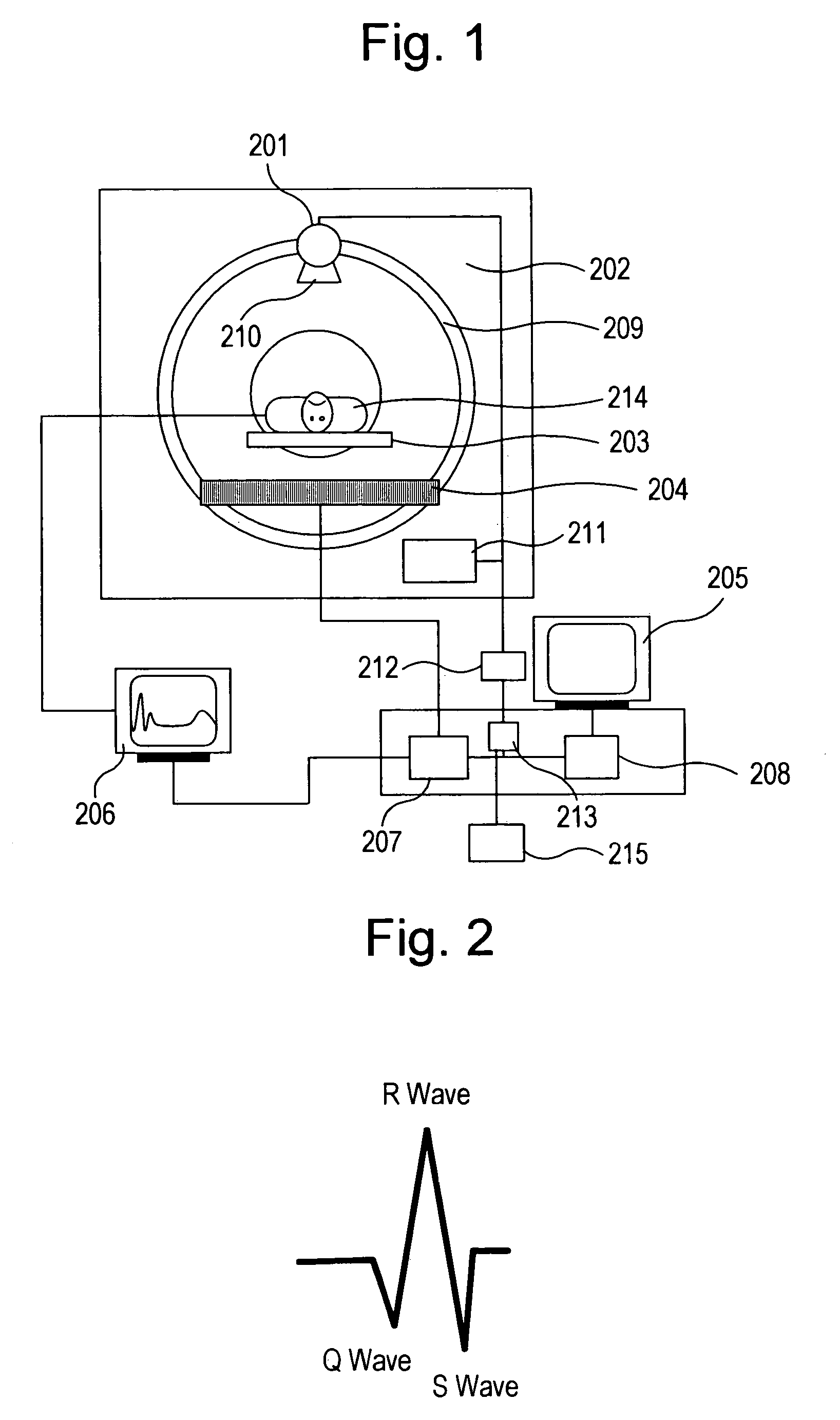
Figure 2 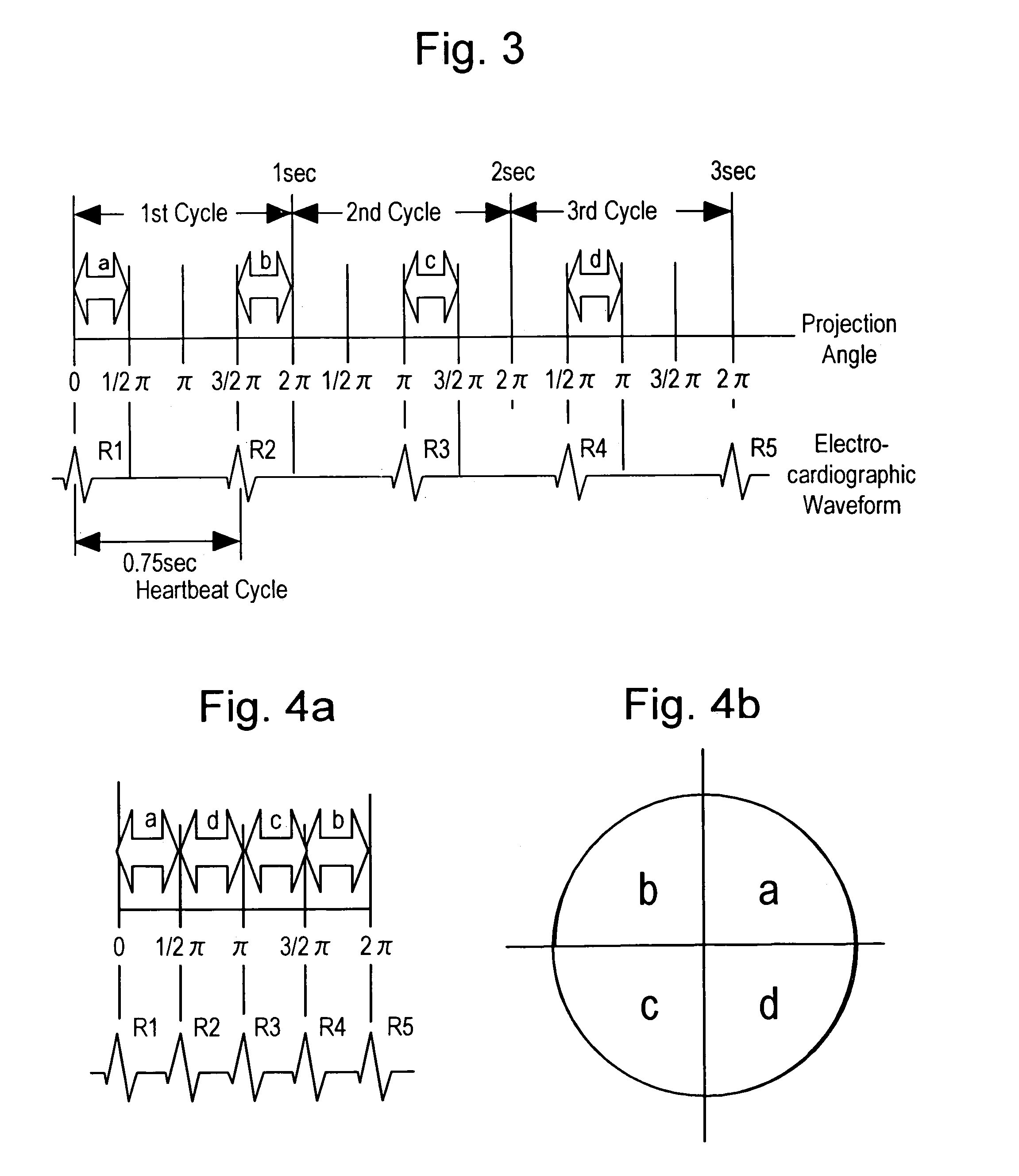
Figure 3
Abstract
Description
TECHNICAL FIELD
[0001] The present invention relates to a method of producing a cardiac tomogram and tomograph using an X-ray CT apparatus, more particularly to a method and apparatus for creating a cardiac tomogram by applying a retrospective ECG (electrocardiography) gating imaging method to an X-ray CT apparatus which performs helical scan and detects projection data with a multi-slice detector.BACKGROUND OF THE INVENTION
[0002] Generally, when a heart of human body is imaged by an X-ray CT apparatus, the image is diagnostically undesirable when motion artifacts appear on the tomogram due to its heartbeat. To prevent motion artifacts from being generated, it is necessary to highly speed up scanning over one heartbeat cycle to a speed of one heartbeat cycle or less. However, heart rates depend on individual bodies, and physical and mental conditions at the point of imaging in the same body. With a scanner mechanism of the current third-generation CT system, it is hard to highly raise ...
Examples
first embodiment
[0052]A method of producing a cardiac tomogram and tomograph using an X-ray CT apparatus according to the present invention will be described, in which a cardiac tomogram having few artifacts caused by discontinuity of projection data can be obtained when the retrospective ECG gating imaging method is applied to helical scan in which the table moves.
(1) In the Case Where the Discontinuous Portion of Projection Data is Narrow
[0053]FIG. 6 shows a case where the multi-slice detector having four lines is used, a scan cycle is 1 second, a heartbeat cycle is 0.75 seconds, and the discontinuous portion of projection data is narrow.
[0054]In the figure, the ordinate axis designates a linear movement direction of a table on which the object to be examined is laid (longitudinal direction of the object, also referred to as Z-axis direction), and the abscissa axis designates a projection angle. Trajectory of the detector traces the center of each detector. The helical scan pitch is “pitch 1” in ...
second embodiment
[0070]Next, a method of producing a cardiac tomogram and tomograph according to the present invention will be described, in which a cardiac tomogram (two-dimensional image), a three-dimensional image, and a four-dimensional image (moving image) can be obtained by optimizing projection data in correspondence with phase difference between a heartbeat cycle and a scan cycle when projection data of the continuous portion obtained by the present invention described above, i.e., projection data at a matched heartbeat time phase are collected in each scan cycle.
[0071]Meanwhile, the X-ray detector 204 used for creating a cardiac tomogram according to the invention is a multi-slice detector in which a plurality of detector elements are aligned in the longitudinal direction of an object to be examined so as to acquire projection data of a plurality of positions at once, and the scan system here is the helical scan.
[0072]As described in FIG. 1, X-rays are radiated from the X-ray tube 201 while...