
Biopsy device handle assembly
a technology for biopsy devices and handle assemblies, which is applied in medical science, surgery, and vaccination/ovulation diagnostics. it can solve problems such as difficult balance of attributes, and achieve the effects of facilitating control and accuracy of the biopsy device, enhancing comfort and maneuverability for users, and facilitating control and accuracy
- Summary
- Abstract
- Description
- Claims
- Application Information
AI Technical Summary
Benefits of technology
Problems solved by technology
Method used
Image

Examples
example 1
Bone Biopsy Core Sample Procedure
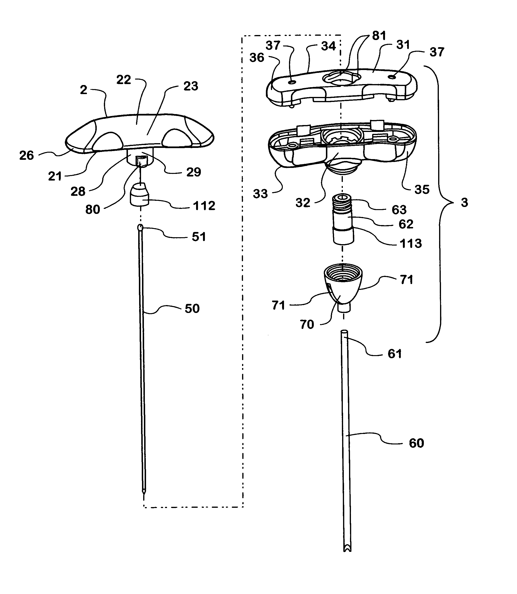
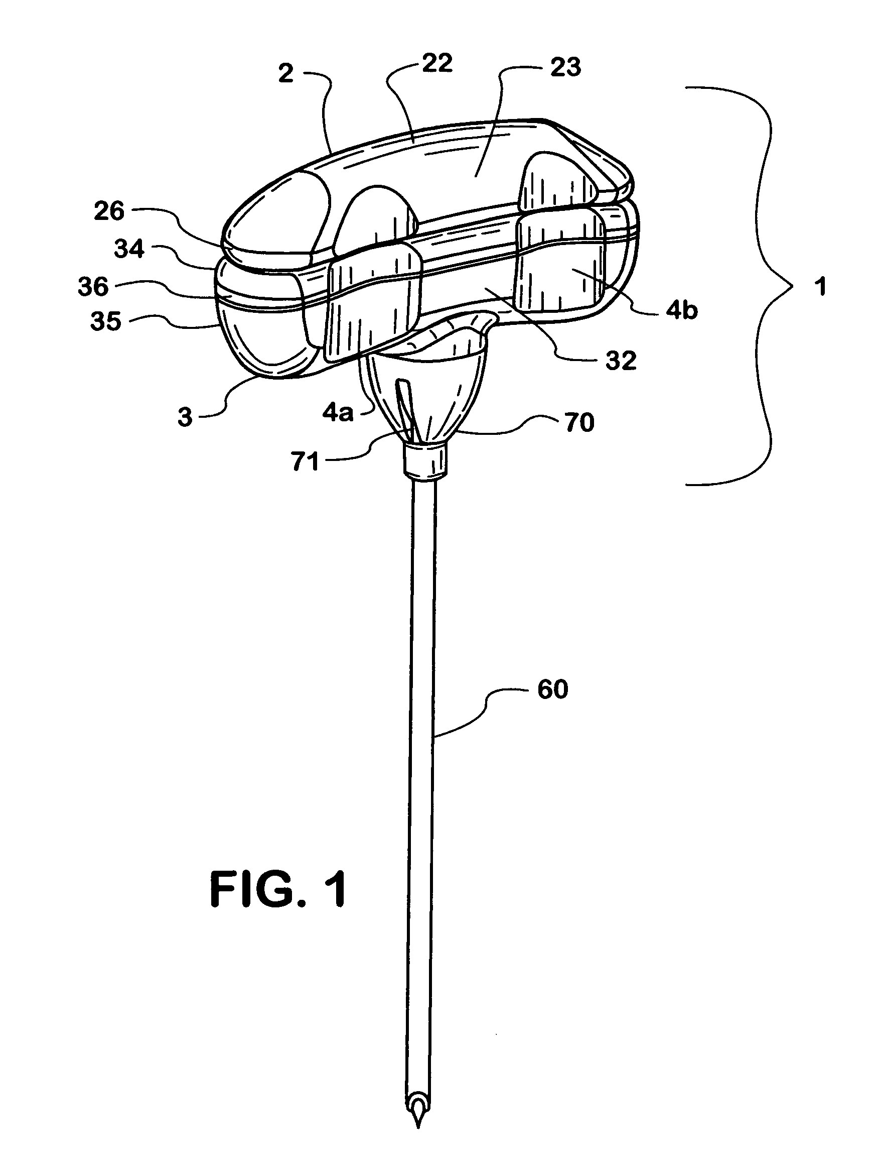
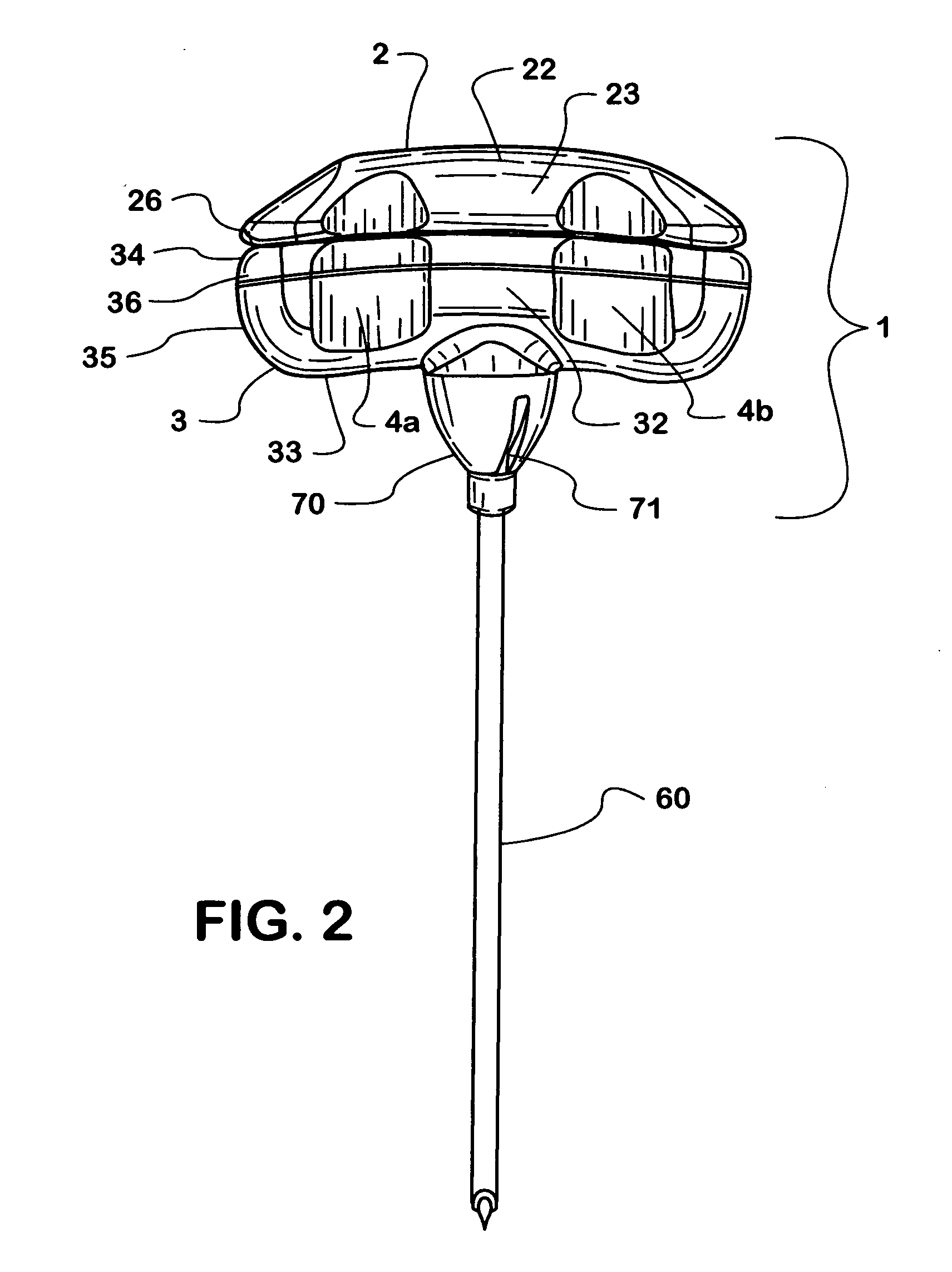
[0052] The bone biopsy core sampling device is generally structured with an inner stylet which functions as a trocar and an outer cannula with a sharpened distal tip to facilitate penetration through the bone tissue to obtain the core tissue sample. Once the sampling site has been determined, the assembled device with the handle portions coupled is forced through the tissue until the distal portion of the outer cannula containing the inner stylet reaches the sampling area. At this point, the inner stylet coupled to the top portion of the handle can be removed and the outer cannula further advanced into the tissue by virtue of force exerted by the user on the handle both longitudinally and rotationally. The user can then determine or estimate the sample length by reinsertion of the inner stylet and viewing the extent that the proximal end of the inner stylet extends beyond the upper surface of the bottom portion of the handle assembly. This step can ...
example 2
Bone Marrow Aspiration Procedure
[0053] The bone marrow aspiration device is generally structured with an inner stylet which functions as a trocar and an outer cannula which functions as an aspiration cannula when a suction source, such as a syringe, is attached to the proximal end thereof. Once the sampling site has been selected and an anatomical assessment made of the patient's bone geometry and dimensions, the adjustable depth guide assembly is adjusted to the appropriate length. The assembled device with the handle portions coupled is then forced through the tissue until the distal portion of the outer cannula containing the inner stylet reaches the sampling area. Ideally, the distal tip of the outer cannula is positioned within the bone at the location to permits unobstructed fluid ingress into the outer cannula. At this point, the inner stylet coupled to the top portion of the handle can be removed, and an aspiration source such as a syringe is then attached to the proximal h...
PUM
 Abstract
Description
Claims
Application Information