

Method for detecting target substance utilizing probe desorption
Patent Information
- Authority / Receiving Office
- US · United States
- Current Assignee / Owner
- Publication Date
- 2008-09-11
Smart Images
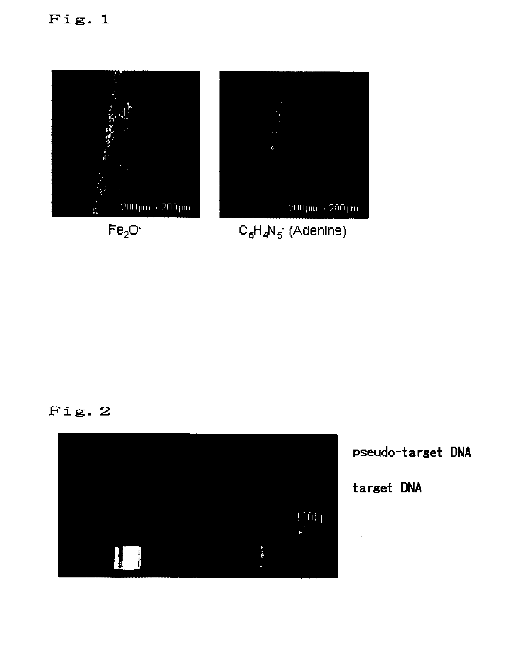
Figure 1 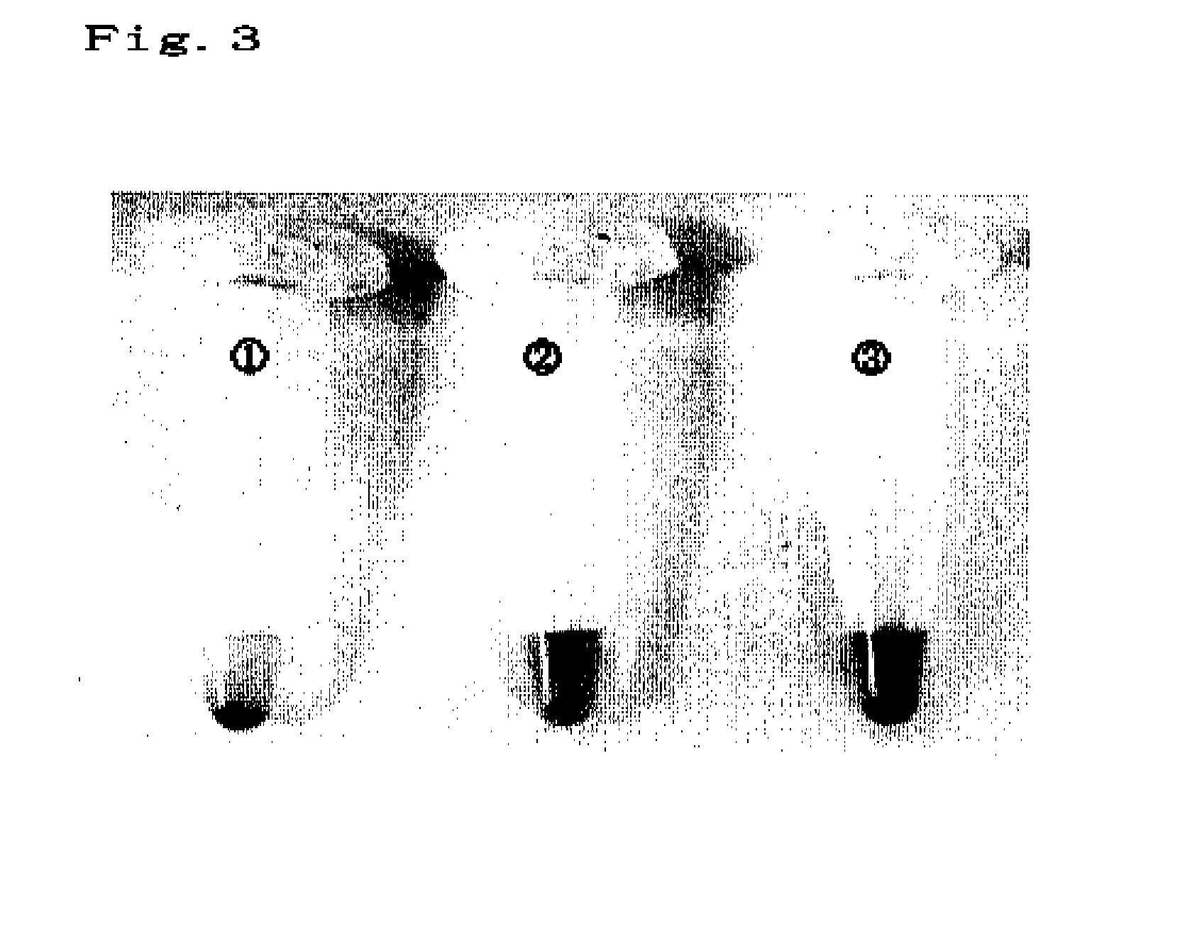
Figure 2 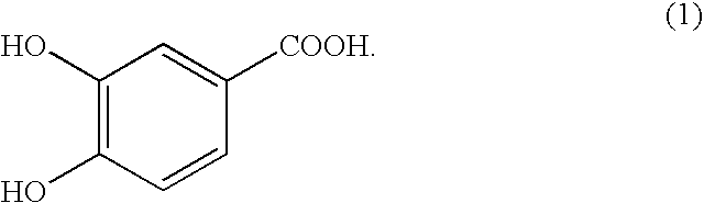
Figure 3
Abstract
Description
TECHNICAL FIELD
[0001] The present invention relates to a method for detecting a target substance with the use of a complex of a fine particle and a probe, wherein desorption of a probe from a fine particle is utilized.BACKGROUND ART
[0002] Methods for detecting biomarkers (e.g., nucleic acids, proteins, lipids, sugar chains, and hormones) are very important for analytical diagnosis. In particular, detection methods with the use of fine particles have been gaining attention because high reaction efficiency (due to the large specific surface area) and high rates of reaction (due to motility greater than that of a bulk body) are expected to be obtained thereby.
[0003] An example of a method for detecting a specimen with the use of a fine particle is described in JP Patent Publication (Kohyo) No. 2004-501340 A. This method is a method for detecting a specimen with the use of a gold nanoparticle to which an oligonucleotide has adhered. The method involves a technique for detecting color chang...
Examples
example 1
Production of Magnetic Particles
[0036]Ferrous acetate (II) (15.66 g) was dissolved in a mixture of ethanol (600 ml) and water (9 ml), followed by heating at 75° C. The obtained solution was mixed with ethanol (300 ml) containing dissolved potassium hydroxide (6.9 g), and further heated at 75° C. for 2 hours. Thereafter, the resulting solution was heated to 100° C. and concentrated at a volume of 300 ml (for approximately 2 hours). After being allowed to stand at 75° C. for 5 hours, the resultant was cooled to room temperature. As a result of the above operation, iron oxide particles dispersed in ethanol were obtained.
example 2
Coating with a Dispersant
[0037]Water (70 ml) was added to ethanol containing dispersed iron oxide particles (30 ml) obtained in Example 1 for precipitation of iron oxide particles. Then, the supernatant was removed therefrom, followed by washing. Subsequently, a compound (dispersant) represented by the following formula (I) was dispersed in water (60 ml):
Then, the obtained solution was added to washed iron oxide particles such that the compound was added at a weight that was 20% of that of iron oxide. Ultrasonication was carried out for an hour, followed by overnight agitation at room temperature. Accordingly, water-dispersible magnetic particles were obtained.
example 3
Immobilization of Nucleic Acid by Adsorption
[0038]The water-dispersible magnetic particles obtained in Example 2, DNA having the sequence represented by SEQ ID NO: 1, and MES buffer were mixed and adjusted to final concentrations of 1 g / L, 50 μM, and 0.1 M, respectively. The resulting mixture was shaken overnight and repeatedly subjected to centrifugation, removal of the supernatant, and redispersion. Thus, unimmobilized DNAs were removed. (5′)-GGGCATOGGTCAGAAGGATT-(3′) (SEQ ID NO: 1)