
Treatment of fibrotic conditions
a fibrotic condition and treatment technology, applied in the field of fibrotic conditions, can solve the problems of unsatisfactory current treatment of keloid scars and excessive respons
Patent Information
- Authority / Receiving Office
- US · United States
- Patent Type
- Applications(United States)
- Current Assignee / Owner
- Publication Date
- 2019-02-14
- Estimated Expiration
- Not applicable · inactive patent
Smart Images
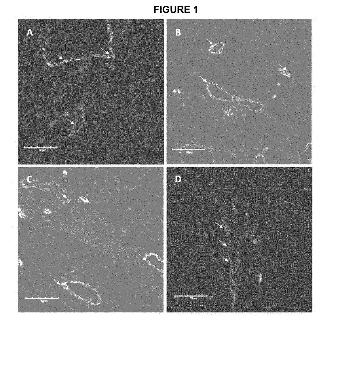
Figure 1 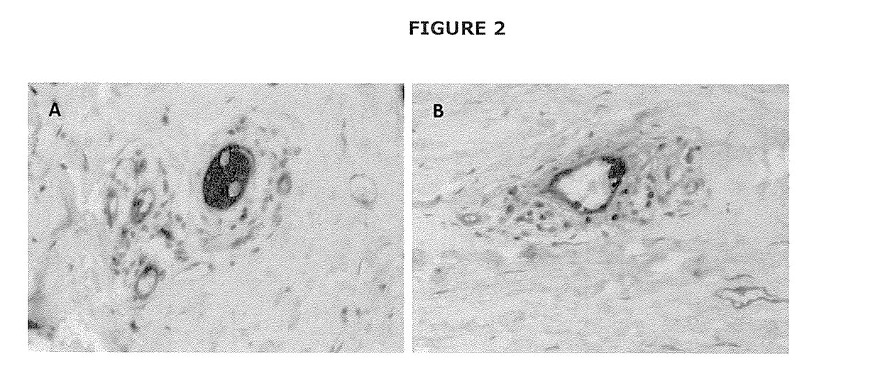
Figure 2 
Figure 3
Abstract
Description
TECHNICAL FIELD
[0001] The present invention provides novel approaches to the treatment of fibrotic conditions. In particular, the present invention provides identification of embryonic stem cell populations shown to be associated with fibrotic lesions, which provide a unique therapeutic target in the treatment of fibrotic conditions.BACKGROUND OF THE INVENTION
[0002] Fibrosis is the formation of excess fibrous connective tissue in an organ or tissue in a reparative or reactive process. This can be a reactive, benign, or pathological state. In response to injury, this is called scarring, and if fibrosis arises from a single cell line, this is called a fibroma. Physiologically, fibrosis acts to deposit connective tissue, which can obliterate the architecture and function of the underlying organ or tissue. Fibrosis can be used to describe the pathological state of excess deposition of fibrous tissue, as well as the process of connective tissue deposition in healing.
[0003] Fibrosis is simil...
Examples
example 1
and Methods
Tissue Samples and Patient Characteristics
[0140]Formalin-fixed, paraffin-embedded keloid scar samples with matching snap-frozen tissues obtained from 3 female and 3 male patients, with a mean age of 29 (range: 22 to 55) years, were used in this study.
[0141]Immunohistochemical (IHC) staining was performed on 5 μm-thick formalin-fixed paraffin-embedded sections of keloid scar samples from 6 patients, according to a protocol approved by the Central Regional Health and Disability Ethics Committee. Antigen retrieval was performed using sodium citrate (Leica, Sydney, Australia) at 95° C. for 15 min, followed by 3,3 diaminobenzidine (DAB) staining with primary antibodies: SOX2, 1:500 (Thermofisher Scientific, CA, USA); pSTAT3, 1:100 (Cell Signalling Technology, MA, USA); OCT4, 1:30 (Cell Marque, CA, USA); NANOG, 1:1000 (Cell Signalling Technology); von Willebrand factor (vWF); 1:200 (Dako, Glostrup, Denmark); tryptase; ready-to-use (Leica) using the bond poly...
example 2
Immunohistochemical Staining
[0146]To investigate the expression of ESC markers in keloid scar Applicants initially examined the expression of OCT4 (FIG. 1A, green), which was localised to the endothelium of the microvessels that expressed vWF (FIG. 1A, red). To further characterise this ESC population, Applicants examined the expression of SOX2 (FIG. 1B, red), involved in ESC gene support (Itinteang, 2012), which was also expressed on the CD34+ endothelium (FIG. 1B, green). pSTAT3 (FIG. 1C, red), the activated form of STAT3, has been associated with ESC self-renewal (Niwa, 1998), was also expressed on the CD34+ endothelium (FIG. 1C, green). NANOG (FIG. 1D, red), another ESC marker which is associated with ESC pluripotency (Loh, 2006), was also expressed on the CD34+ endothelium (FIG. 1D, green). The abundance of nucleated cells (FIG. 1A-D) immediately adjacent to the endothelium that expressed these ESC markers, appeared similar to a recent report of KALTs in keloid scar and led App...
example 3
n
[0148]The presence of T cells, B cells, mast cells and M2 macrophages has been recently demonstrated in keloid scars and has been referred to as KALTs (Bagabir, 2012). One report describes the effect of IL6 / IL17 in the development of keloid scar (Zhang, 2009).
[0149]Applicant's results highlight the intriguing novel finding of the expression of the ESC markers OCT4, SOX2, NANOG and pSTAT3 on the endothelium of the microvessels surrounded by the KALTs which are located just beneath the epidermis (Bagabir, 2012).
[0150]It is exciting to speculate that this primitive endothelium within keloid scar may potentially be the source of both myofibroblasts and KALTs, although this remains to be conclusively determined.
[0151]ESCs are undifferentiated cells possessing the ability to differentiate and proliferate indefinitely (Bishop, 2002). They may be induced to differentiate into a broad range of cell types, with vastly different properties (Bishop, 2002). The fate of stem cells within tissues...