
Patient stratification for cancer therapy based on genomic DNA microarray analysis
Patent Information
- Authority / Receiving Office
- US · United States
- Current Assignee / Owner
- Publication Date
- 2017-01-03
- Estimated Expiration
- Not applicable · inactive patent
Smart Images
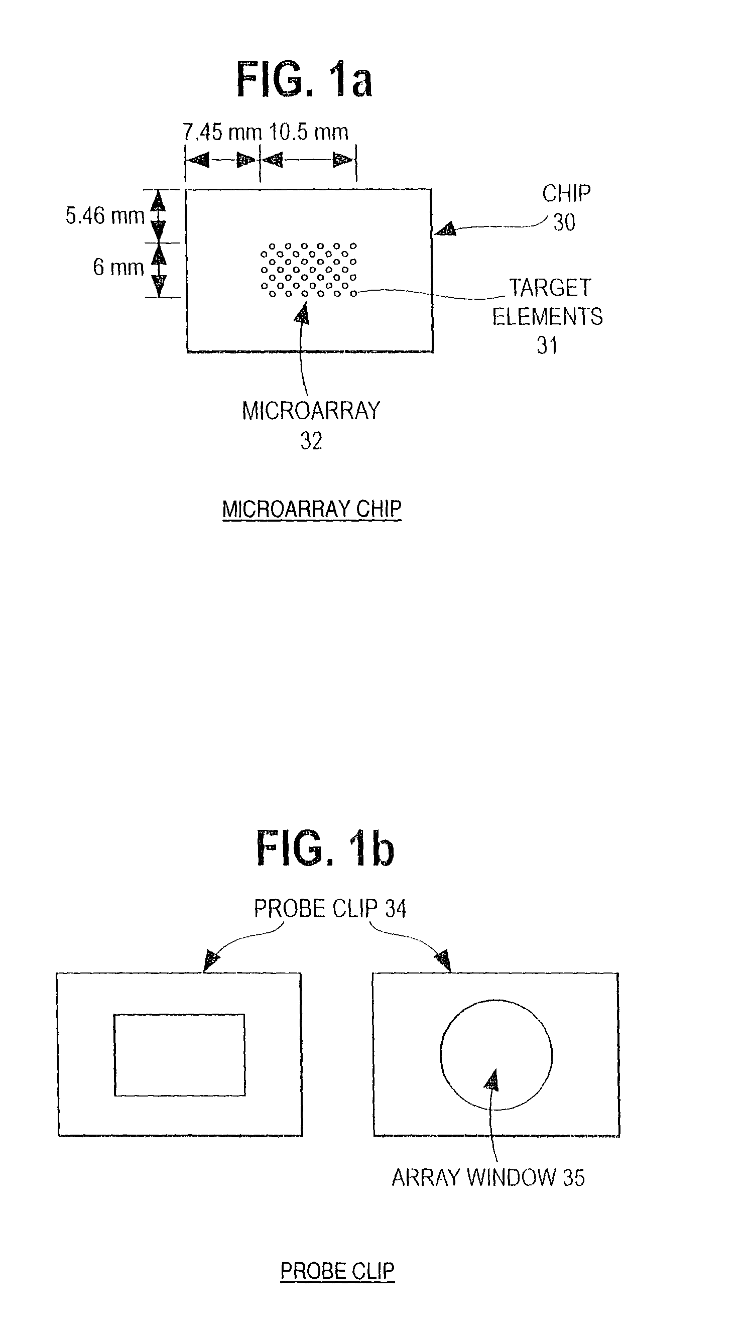
Figure 1 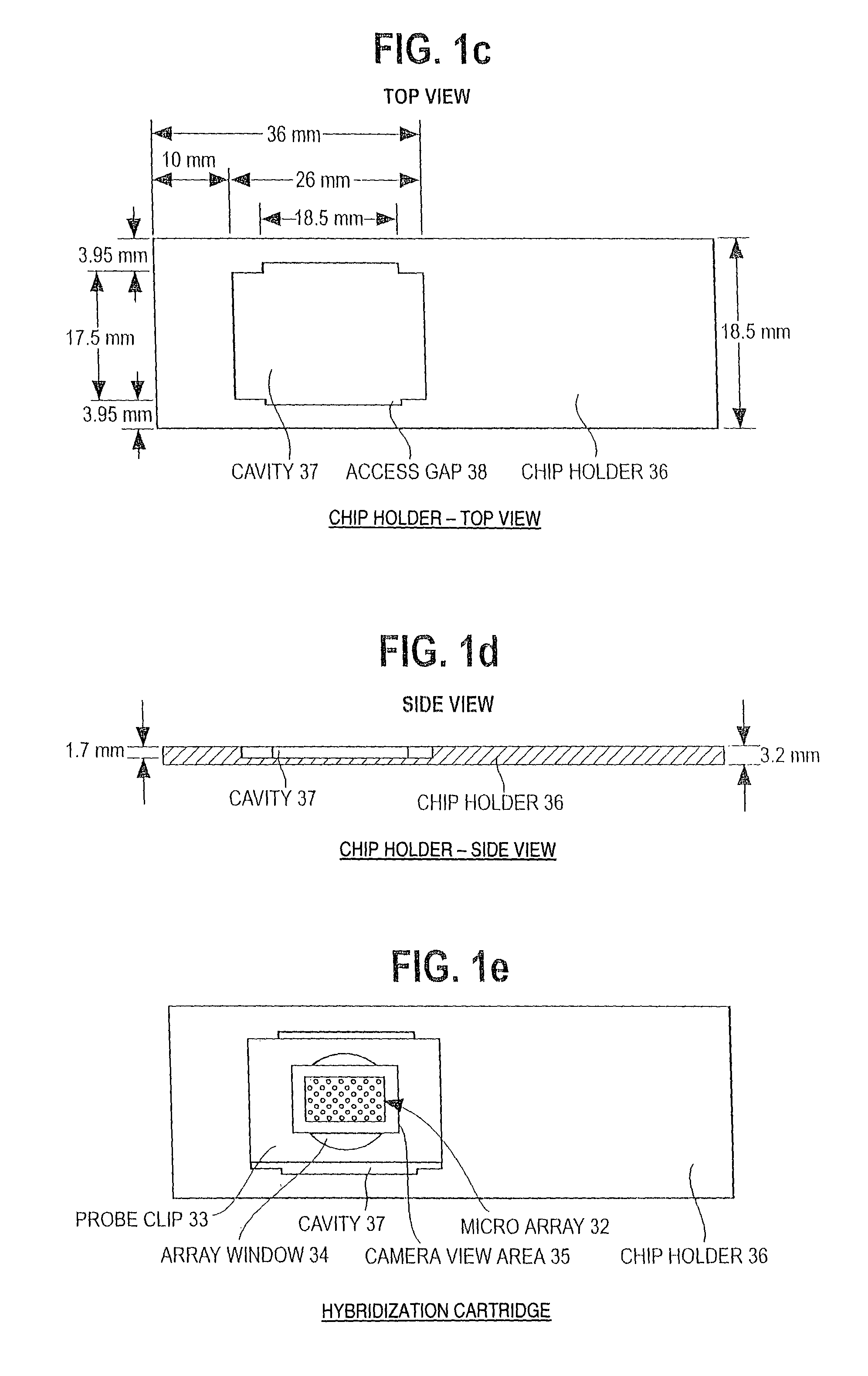
Figure 2
Abstract
Description
RELATED APPLICATION
[0001] This application is a continuation-in-part of commonly Assigned U.S. patent application Ser. No. 09 / 243,067, filed 2 Feb. 1999, “Simultaneous Measurement of Gene Expression and Genomic Abnormalities Using Nucleic Acid Microarrays, Y. Bao, et al, now U.S. Pat. No. 6,251,601 B1”.FIELD OF THE INVENTION
[0002] This invention relates generally to the use of nucleic acid probe test to guide therapeutic selection for the treatment of cancer. More particularly, the invention relates to the stratification of a human cancer patient population into various cancer therapy groups based on the use of a genomic deoxyribonucleic acid (“DNA”) microarray to assess a tissue sample from the patient.BACKGROUND OF THE INVENTION
[0003] Abnormalities in the expression of genes, both in the timing and level of expression of particular genes, are a fundamental cause of cancer and other human disease. Abnormalities in genomic DNA, i.e. in chromosomes, are also a fundamental cause of cance...
Examples
example 1
(A) Procedures
[0137](i) Microdissected breast cancer tissue extracts were obtained from Dr. John Bartlett, Glasgow Royal Infirmary, University of Glasgow. These extracts were produced by first staining thawed, previously frozen breast cancer tissue sections with toluidine blue. Tumor / normal tissue was identified under a ×50 dissecting microscope, followed by microdissection with a scalpel blade into a microfuge tube. One sample (#10) was classified as normal tissue adjacent to tumor tissue (#9). The chromosomal DNA was extracted from the microdissected samples by addition of 25 microliters of proteinase K in TE buffer (pH 7.4) and incubation at 37 degrees C. for 48 hours. The proteinase K was then denatured by heading at 95 degrees C. for 20 minutes.
[0138]5 microliters of each 25 microliter of the proteinase K digested sample was amplified using DOP-PCR as described in Speicher, M. R. et al. (1993) Human Molecular Genetics 2, 1907-1914, using as a primer:
[0139]5′-CCG ACT CGA GNN NNN...