How does P wave dispersion affect atrial conduction
AUG 19, 20259 MIN READ
Generate Your Research Report Instantly with AI Agent
Patsnap Eureka helps you evaluate technical feasibility & market potential.
P Wave Dispersion Background and Objectives
P wave dispersion is a crucial electrophysiological phenomenon that has gained significant attention in the field of cardiology over the past few decades. This concept refers to the variation in P wave duration across different leads of a standard 12-lead electrocardiogram (ECG). The P wave, representing atrial depolarization, serves as a critical indicator of atrial conduction and has been increasingly recognized for its potential in predicting various cardiac arrhythmias, particularly atrial fibrillation.
The study of P wave dispersion emerged in the late 1990s as researchers sought more refined methods to assess atrial electrical activity. Initially, it was observed that patients with a history of paroxysmal atrial fibrillation exhibited greater P wave dispersion compared to healthy individuals. This discovery sparked a surge of interest in exploring the relationship between P wave dispersion and atrial conduction abnormalities.
As research in this area progressed, it became evident that P wave dispersion could provide valuable insights into the electrophysiological properties of the atria. The primary objective of studying P wave dispersion is to understand its role in atrial conduction and its potential as a predictor of atrial arrhythmias. By analyzing the variability in P wave duration across different ECG leads, researchers aim to detect subtle changes in atrial conduction that may precede the onset of clinically significant arrhythmias.
The technological advancements in ECG recording and analysis have significantly contributed to the evolution of P wave dispersion studies. High-resolution ECG systems and sophisticated signal processing algorithms have enabled more accurate measurements of P wave parameters, including duration and morphology. These developments have paved the way for more precise quantification of P wave dispersion and its correlation with atrial conduction properties.
Current research objectives in the field of P wave dispersion focus on several key areas. Firstly, there is a concerted effort to establish standardized measurement techniques and reference values for P wave dispersion across different populations. This standardization is crucial for the widespread clinical application of P wave dispersion analysis. Secondly, researchers are investigating the underlying mechanisms by which increased P wave dispersion affects atrial conduction, exploring factors such as atrial fibrosis, electrical remodeling, and autonomic nervous system influences.
Furthermore, there is growing interest in utilizing P wave dispersion as a risk stratification tool for various cardiovascular conditions. Studies are being conducted to evaluate its predictive value not only for atrial fibrillation but also for other cardiac disorders such as hypertension, heart failure, and coronary artery disease. The ultimate goal is to develop P wave dispersion into a reliable, non-invasive marker for early detection of atrial conduction abnormalities and risk assessment of atrial arrhythmias.
Clinical Relevance of P Wave Dispersion Analysis
P wave dispersion analysis has emerged as a significant tool in clinical cardiology, offering valuable insights into atrial conduction abnormalities and potential arrhythmogenic risks. This non-invasive electrocardiographic parameter has gained considerable attention due to its potential to predict and assess various cardiac conditions, particularly atrial fibrillation.
The clinical relevance of P wave dispersion analysis lies primarily in its ability to detect subtle changes in atrial conduction that may precede the onset of atrial arrhythmias. By measuring the difference between the longest and shortest P wave durations in a 12-lead ECG, clinicians can assess the heterogeneity of atrial depolarization. Increased P wave dispersion has been associated with a higher risk of developing atrial fibrillation, making it a useful marker for identifying patients who may benefit from closer monitoring or preventive interventions.
In the context of atrial fibrillation management, P wave dispersion analysis has shown promise in predicting the recurrence of arrhythmia following cardioversion or ablation procedures. This information can guide clinicians in tailoring treatment strategies and follow-up protocols for individual patients, potentially improving long-term outcomes and reducing the burden of repeated interventions.
Beyond atrial fibrillation, P wave dispersion analysis has demonstrated clinical utility in various cardiovascular conditions. Studies have shown its potential in assessing the risk of atrial arrhythmias in patients with hypertension, coronary artery disease, and heart failure. Additionally, it has been investigated as a prognostic marker in non-cardiac conditions such as chronic obstructive pulmonary disease and obstructive sleep apnea, where altered atrial conduction may contribute to cardiovascular complications.
The integration of P wave dispersion analysis into routine clinical practice offers several advantages. It is a simple, cost-effective, and readily available method that can be performed using standard ECG equipment. This accessibility makes it an attractive option for risk stratification in both primary care and specialized cardiology settings. Furthermore, the non-invasive nature of the technique allows for repeated measurements over time, enabling clinicians to monitor changes in atrial conduction and adjust management strategies accordingly.
However, it is important to note that while P wave dispersion analysis shows promise, its clinical application still faces some challenges. Standardization of measurement techniques and establishment of universally accepted reference values are ongoing areas of research. Additionally, the predictive value of P wave dispersion may vary depending on the specific clinical context and patient population, necessitating careful interpretation in conjunction with other clinical and diagnostic parameters.
Current Challenges in P Wave Dispersion Assessment
P wave dispersion assessment faces several significant challenges that hinder its widespread clinical application and reliability. One of the primary obstacles is the lack of standardization in measurement techniques. Different studies and clinical settings employ varying methodologies for measuring P wave dispersion, leading to inconsistent results and difficulties in comparing findings across research efforts.
The manual measurement of P wave dispersion is another major challenge. This process is time-consuming and prone to human error, particularly when dealing with large volumes of ECG data. The subjectivity involved in manual measurements can lead to inter-observer and intra-observer variability, compromising the reproducibility of results.
Technical limitations of ECG recording systems also contribute to the challenges in P wave dispersion assessment. Many standard ECG machines have limited sampling rates and inadequate resolution, which can affect the accurate detection of subtle changes in P wave morphology. This technical constraint may result in underestimation or overestimation of P wave dispersion values.
The influence of physiological and pathological factors on P wave morphology further complicates the assessment process. Factors such as age, heart rate, autonomic tone, and underlying cardiac conditions can all affect P wave characteristics, making it difficult to establish universally applicable normal ranges and cut-off values for clinical decision-making.
Another significant challenge lies in the interpretation of P wave dispersion values in the context of atrial conduction abnormalities. While increased P wave dispersion has been associated with various atrial pathologies, the specificity and sensitivity of this parameter for predicting specific conduction disorders or arrhythmias remain subjects of ongoing debate.
The lack of large-scale, prospective studies validating the prognostic value of P wave dispersion in different clinical scenarios poses a challenge to its widespread adoption. This gap in evidence limits the confidence of clinicians in using P wave dispersion as a reliable marker for risk stratification or treatment guidance.
Furthermore, the integration of P wave dispersion assessment into routine clinical practice faces practical hurdles. Many healthcare settings lack the necessary software tools or expertise to perform automated P wave dispersion measurements, limiting its accessibility and utility in everyday patient care.
Existing Methods for Measuring P Wave Dispersion
01 P wave dispersion measurement and analysis
Methods and systems for measuring and analyzing P wave dispersion in electrocardiogram (ECG) signals. This involves detecting and quantifying variations in P wave morphology across different ECG leads, which can provide insights into atrial conduction heterogeneity and potential arrhythmia risk.- P-wave dispersion measurement and analysis: Methods and systems for measuring and analyzing P-wave dispersion in electrocardiogram (ECG) signals to assess atrial conduction and predict atrial fibrillation risk. This involves detecting P-waves, calculating dispersion parameters, and using algorithms to interpret the results for clinical diagnosis and treatment planning.
- Atrial pacing techniques for improved conduction: Cardiac pacing methods and devices designed to optimize atrial conduction and reduce P-wave dispersion. These techniques may involve multi-site atrial pacing, adaptive pacing algorithms, or specialized electrode configurations to improve synchronization of atrial activation and reduce the risk of atrial arrhythmias.
- Non-invasive atrial conduction assessment: Non-invasive methods for evaluating atrial conduction and P-wave characteristics using advanced signal processing of surface ECG or other external sensors. These techniques aim to provide detailed information about atrial electrical activity without the need for invasive procedures.
- Implantable devices for monitoring atrial conduction: Implantable cardiac devices equipped with specialized sensors and algorithms to continuously monitor atrial conduction patterns, P-wave dispersion, and other relevant parameters. These devices can provide long-term data for assessing arrhythmia risk and guiding treatment decisions.
- Atrial conduction modeling and simulation: Computational methods and systems for modeling and simulating atrial conduction patterns, including P-wave propagation and dispersion. These models can be used to predict arrhythmia risk, optimize treatment strategies, and enhance understanding of atrial electrophysiology.
02 Atrial conduction time assessment
Techniques for assessing atrial conduction time using various cardiac signals. This includes methods for measuring inter-atrial conduction delays and identifying conduction abnormalities that may contribute to atrial arrhythmias.Expand Specific Solutions03 Cardiac pacing optimization based on atrial conduction
Approaches for optimizing cardiac pacing parameters based on atrial conduction characteristics. This involves adjusting pacing timing and modes to compensate for atrial conduction delays and improve overall cardiac synchrony.Expand Specific Solutions04 Atrial fibrillation risk assessment
Methods for assessing the risk of atrial fibrillation using P wave dispersion and other atrial conduction parameters. This includes developing predictive models and risk stratification tools based on ECG and other cardiac signal analyses.Expand Specific Solutions05 Non-invasive atrial conduction mapping
Techniques for non-invasive mapping of atrial conduction patterns using advanced signal processing of surface ECG and other non-invasive cardiac signals. This allows for detailed characterization of atrial activation sequences without the need for invasive procedures.Expand Specific Solutions
Key Researchers and Institutions in Cardiac Electrophysiology
The competitive landscape for P wave dispersion and atrial conduction technology is in a growth phase, with increasing market size and advancing technological maturity. The market is driven by the rising prevalence of cardiovascular diseases and the growing demand for non-invasive diagnostic tools. Key players like Medtronic, BIOTRONIK, and Abbott (through Pacesetter) are leading the field with innovative ECG and cardiac monitoring devices. Emerging companies such as Bardy Diagnostics and Biosense Webster are also making significant contributions, focusing on P-wave centric ECG detection and advanced mapping technologies. Academic institutions like King's College London and the University of Freiburg are conducting cutting-edge research, further advancing the understanding of P wave dispersion and its clinical implications.
Medtronic, Inc.
Technical Solution: Medtronic has developed advanced cardiac mapping systems that analyze P wave dispersion to assess atrial conduction. Their EnSite Precision cardiac mapping system uses high-density electroanatomical mapping to create detailed 3D models of the atria, allowing for precise visualization of P wave propagation and dispersion patterns[1]. This technology enables electrophysiologists to identify areas of abnormal conduction and potential arrhythmogenic substrates. Medtronic has also integrated P wave dispersion analysis into their implantable cardiac monitors and pacemakers, using proprietary algorithms to detect subtle changes in atrial conduction that may precede atrial fibrillation[2].
Strengths: Comprehensive cardiac mapping capabilities, integration of P wave analysis in implantable devices. Weaknesses: Complexity and cost of advanced mapping systems may limit widespread adoption.
Biosense Webster (Israel) Ltd.
Technical Solution: Biosense Webster has developed the CARTO 3 System, an advanced 3D electroanatomical mapping platform that incorporates P wave analysis for atrial conduction assessment. The system uses proprietary algorithms to analyze P wave morphology and dispersion across multiple leads, providing a detailed map of atrial activation patterns[3]. Their CONFIDENSE Module enhances the resolution of atrial mapping, allowing for precise localization of areas with abnormal conduction. Biosense Webster has also introduced the QMODE+ mapping feature, which enables rapid, high-density mapping of P wave characteristics during sinus rhythm, facilitating the identification of potential arrhythmia substrates[4].
Strengths: High-resolution 3D mapping, advanced P wave analysis algorithms. Weaknesses: System complexity may require specialized training for optimal use.
Innovative Approaches in P Wave Dispersion Quantification
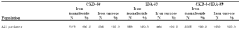
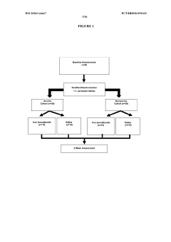
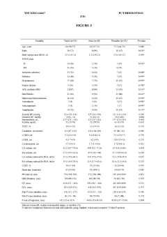
Treating iron deficiency in subjects at risk of cardiovascular adverse events and iron for the management of atrial fibrillation
PatentWO2020144667A1
Innovation
- Administering iron isomaltoside, a high-dose intravenous iron carbohydrate complex, to patients at risk of cardiovascular adverse events, including those with a history of myocardial infarction, stroke, atrial fibrillation, congestive heart failure, and chronic kidney disease, to improve iron stores and reduce cardiovascular risk, combined with other medications to enhance therapeutic benefits.
Implications for Arrhythmia Risk Stratification
P wave dispersion, a measure of atrial conduction heterogeneity, has significant implications for arrhythmia risk stratification. This parameter, derived from surface electrocardiograms, provides valuable insights into the electrical properties of the atria and their potential for arrhythmogenesis.
Increased P wave dispersion is associated with a higher risk of atrial fibrillation (AF), the most common sustained cardiac arrhythmia. By quantifying the variability in atrial conduction, P wave dispersion serves as a non-invasive marker for identifying patients at elevated risk for AF development or recurrence. This information is particularly crucial in guiding preventive strategies and treatment decisions for high-risk individuals.
Furthermore, P wave dispersion has demonstrated prognostic value in various cardiovascular conditions. In patients with heart failure, increased P wave dispersion correlates with a higher incidence of atrial arrhythmias and adverse cardiovascular events. Similarly, in hypertensive patients, elevated P wave dispersion may indicate subclinical atrial remodeling and increased susceptibility to arrhythmias, allowing for early intervention and risk modification.
The utility of P wave dispersion extends to post-operative risk assessment. Following cardiac surgery, patients with greater P wave dispersion are more likely to develop post-operative AF, a common complication associated with increased morbidity and prolonged hospital stays. By identifying these high-risk patients, clinicians can implement targeted prophylactic measures to reduce the incidence of post-operative arrhythmias.
In the context of structural heart disease, P wave dispersion provides additional information on the arrhythmogenic substrate. Patients with mitral valve disease or left atrial enlargement often exhibit increased P wave dispersion, reflecting the impact of structural remodeling on atrial conduction properties. This knowledge aids in risk stratification and guides decisions regarding the timing of interventions or the need for closer monitoring.
The integration of P wave dispersion into comprehensive risk assessment models enhances the accuracy of arrhythmia prediction. When combined with other electrocardiographic parameters, echocardiographic findings, and clinical risk factors, P wave dispersion contributes to a more nuanced evaluation of arrhythmia risk. This multifaceted approach allows for better patient stratification and personalized management strategies.
As research in this field progresses, the role of P wave dispersion in arrhythmia risk stratification continues to evolve. Emerging evidence suggests its potential application in predicting the success of catheter ablation procedures for AF and guiding antiarrhythmic drug selection. These developments underscore the ongoing importance of P wave dispersion as a valuable tool in the assessment and management of patients at risk for atrial arrhythmias.
Standardization of P Wave Dispersion Measurements
The standardization of P wave dispersion measurements is crucial for ensuring consistency and reliability in assessing atrial conduction abnormalities. This process involves establishing uniform protocols for recording, measuring, and interpreting P wave characteristics across different electrocardiographic (ECG) systems and clinical settings.
One of the primary challenges in standardizing P wave dispersion measurements is the variability in ECG recording techniques. Different lead placements, sampling rates, and filtering methods can significantly affect the appearance and duration of P waves. To address this, international cardiology organizations have proposed guidelines for ECG acquisition, emphasizing the importance of using high-resolution digital systems with standardized lead configurations.
The definition of P wave onset and offset points is another critical aspect of standardization. Manual measurements are subject to inter-observer variability, leading to inconsistencies in P wave duration and dispersion calculations. Automated algorithms have been developed to improve precision and reproducibility. These algorithms typically employ signal processing techniques such as wavelet transforms or template matching to identify P wave boundaries more accurately.
Standardization efforts also focus on establishing normative values for P wave dispersion across different populations. Large-scale studies have been conducted to determine reference ranges, taking into account factors such as age, gender, and ethnicity. These normative data are essential for interpreting individual measurements and identifying abnormal atrial conduction patterns.
The choice of leads for P wave dispersion analysis is another area requiring standardization. While some researchers advocate for using all 12 leads, others propose simplified approaches using specific lead combinations. Consensus guidelines recommend a minimum set of leads that provide comprehensive information about atrial depolarization while balancing practicality in clinical settings.
Temporal and beat-to-beat variability in P wave morphology pose additional challenges for standardization. Methods for averaging multiple cardiac cycles and accounting for respiratory variations have been proposed to enhance measurement stability. Some advanced techniques incorporate dynamic analysis of P wave changes over time, providing insights into the temporal aspects of atrial conduction.
Standardization efforts extend to reporting and interpretation of P wave dispersion measurements. Uniform formats for presenting results, including both numerical values and visual representations, have been proposed to facilitate communication among healthcare providers and researchers. Additionally, guidelines for interpreting P wave dispersion in the context of other ECG parameters and clinical information are being developed to improve diagnostic accuracy and clinical decision-making.
Unlock deeper insights with Patsnap Eureka Quick Research — get a full tech report to explore trends and direct your research. Try now!
Generate Your Research Report Instantly with AI Agent
Supercharge your innovation with Patsnap Eureka AI Agent Platform!