P wave examination in cardiac amyloidosis patients
AUG 19, 20259 MIN READ
Generate Your Research Report Instantly with AI Agent
Patsnap Eureka helps you evaluate technical feasibility & market potential.
P Wave Analysis in Cardiac Amyloidosis: Background and Objectives
Cardiac amyloidosis is a rare but serious condition characterized by the abnormal deposition of amyloid proteins in the heart tissue. This accumulation can lead to significant structural and functional changes in the heart, affecting its ability to pump blood effectively. In recent years, there has been growing interest in the analysis of P waves as a potential diagnostic and prognostic tool for cardiac amyloidosis patients.
The P wave, representing atrial depolarization in the electrocardiogram (ECG), has traditionally been overlooked in favor of other ECG components. However, emerging research suggests that P wave examination may provide valuable insights into the early stages of cardiac amyloidosis and its progression. This renewed focus on P wave analysis aligns with the broader trend in cardiology towards more comprehensive and nuanced interpretations of ECG data.
The primary objective of P wave analysis in cardiac amyloidosis is to identify subtle changes in atrial electrical activity that may precede more obvious clinical manifestations. These changes could potentially serve as early markers of disease onset or progression, allowing for earlier intervention and improved patient outcomes. Additionally, P wave analysis may help differentiate cardiac amyloidosis from other forms of cardiomyopathy, addressing a significant diagnostic challenge in clinical practice.
Another key goal of P wave examination is to enhance risk stratification in cardiac amyloidosis patients. By correlating P wave characteristics with clinical outcomes, researchers aim to develop more accurate prognostic models. This could lead to more personalized treatment strategies and better resource allocation in managing this complex disease.
Furthermore, P wave analysis may provide insights into the underlying pathophysiological mechanisms of cardiac amyloidosis. By studying how amyloid deposition affects atrial electrical properties, researchers hope to gain a deeper understanding of disease progression and identify potential targets for therapeutic intervention.
The technological evolution in ECG recording and analysis has played a crucial role in enabling more detailed P wave examinations. Advanced digital ECG systems, coupled with sophisticated signal processing algorithms, now allow for the detection and quantification of subtle P wave abnormalities that were previously undetectable. This technological progress has opened new avenues for research and clinical applications in cardiac amyloidosis.
As we delve deeper into the potential of P wave analysis in cardiac amyloidosis, it is important to consider its integration with other diagnostic modalities. The ultimate aim is to develop a comprehensive approach that combines P wave examination with imaging techniques, biomarkers, and genetic testing to improve the overall management of cardiac amyloidosis patients.
Clinical Demand for P Wave Examination in Amyloidosis
The clinical demand for P wave examination in cardiac amyloidosis patients has been steadily increasing due to its potential diagnostic and prognostic value. Cardiac amyloidosis, a condition characterized by the deposition of abnormal proteins in the heart tissue, can lead to various cardiac complications, including arrhythmias and heart failure. The P wave, representing atrial depolarization, provides crucial information about atrial function and conduction, which are often affected in amyloidosis patients.
P wave examination offers several advantages in the management of cardiac amyloidosis. Firstly, it allows for early detection of atrial abnormalities, which may precede more severe cardiac manifestations. This early identification can lead to timely interventions and improved patient outcomes. Secondly, P wave analysis can help in risk stratification, identifying patients at higher risk of developing atrial fibrillation or other arrhythmias, which are common complications in amyloidosis.
The demand for P wave examination is further driven by its non-invasive nature and cost-effectiveness. Unlike more invasive diagnostic procedures, P wave analysis can be performed through standard electrocardiography (ECG) or more advanced signal-averaged ECG techniques. This accessibility makes it an attractive option for routine monitoring and follow-up of amyloidosis patients.
Recent studies have highlighted the prognostic significance of P wave parameters in cardiac amyloidosis. Abnormalities in P wave duration, amplitude, and morphology have been associated with increased risk of adverse cardiac events and mortality. This growing body of evidence has led to increased interest in incorporating P wave examination into standard clinical protocols for amyloidosis patients.
The integration of artificial intelligence and machine learning algorithms in ECG analysis has further enhanced the clinical utility of P wave examination. These advanced technologies can detect subtle P wave changes that may be overlooked by conventional analysis, potentially improving diagnostic accuracy and risk prediction.
As the prevalence of cardiac amyloidosis continues to rise, particularly in aging populations, the demand for effective diagnostic and monitoring tools is expected to grow. P wave examination, with its ability to provide valuable insights into atrial function and disease progression, is well-positioned to meet this clinical need. Furthermore, ongoing research into novel therapies for amyloidosis may create additional demand for reliable biomarkers of treatment response, a role that P wave parameters could potentially fulfill.
Current Challenges in P Wave Assessment for Amyloidosis
The assessment of P waves in cardiac amyloidosis patients presents several significant challenges that hinder accurate diagnosis and monitoring of the disease progression. One of the primary difficulties lies in the subtle and often ambiguous nature of P wave changes associated with amyloidosis. These alterations can be easily overlooked or misinterpreted, especially in the early stages of the disease when the cardiac involvement is minimal.
The variability in P wave morphology among amyloidosis patients further complicates the assessment process. The heterogeneous nature of amyloid deposition in the atrial myocardium can lead to diverse P wave patterns, making it challenging to establish standardized criteria for diagnosis. This variability also makes it difficult to differentiate amyloidosis-related changes from other cardiac conditions that may affect P wave characteristics.
Another significant challenge is the limited sensitivity and specificity of current P wave analysis techniques in the context of amyloidosis. Traditional electrocardiographic measurements may not be sufficiently sensitive to detect the subtle changes in atrial conduction that occur in the early stages of the disease. This limitation can result in delayed diagnosis and missed opportunities for early intervention.
The influence of comorbidities and concurrent cardiac conditions on P wave characteristics poses an additional challenge. Many amyloidosis patients, particularly those with AL amyloidosis, may have other cardiac abnormalities that can affect P wave morphology, making it difficult to isolate the specific effects of amyloid deposition on atrial conduction.
Technical limitations in P wave recording and analysis also contribute to the challenges in assessment. The low amplitude of P waves, especially in advanced amyloidosis cases, can make accurate measurement and interpretation challenging. Additionally, the lack of standardized protocols for P wave analysis in the context of amyloidosis further complicates the assessment process.
The dynamic nature of amyloidosis progression presents another hurdle in P wave assessment. As the disease evolves, P wave characteristics may change, requiring frequent reassessment and interpretation. This temporal variability makes it challenging to establish reliable prognostic markers based on P wave analysis alone.
Lastly, the integration of P wave assessment with other diagnostic modalities remains a challenge. While P wave analysis can provide valuable insights into atrial function and conduction, its correlation with other imaging and biomarker findings in amyloidosis is not always straightforward. This complexity makes it difficult to develop a comprehensive diagnostic and prognostic approach that effectively incorporates P wave assessment alongside other clinical parameters.
Existing P Wave Examination Methods for Amyloidosis
01 P wave detection and analysis in ECG signals
Methods and systems for detecting and analyzing P waves in electrocardiogram (ECG) signals. This includes techniques for identifying P wave morphology, measuring P wave duration and amplitude, and distinguishing P waves from other ECG components. These approaches can be used for diagnosing various cardiac conditions and assessing atrial function.- P wave detection and analysis in ECG signals: Methods and systems for detecting and analyzing P waves in electrocardiogram (ECG) signals. These techniques involve identifying P wave characteristics, such as amplitude, duration, and morphology, to assess cardiac function and diagnose atrial abnormalities.
- P wave signal processing in communication systems: Techniques for processing P waves in communication systems, including modulation, demodulation, and filtering methods. These approaches aim to improve signal quality, reduce interference, and enhance overall system performance in wireless and wired communication networks.
- P wave analysis for seismic exploration: Methods for analyzing P waves in seismic data to characterize subsurface structures and properties. These techniques involve processing and interpreting P wave information to identify geological features, estimate rock properties, and locate potential hydrocarbon reservoirs.
- P wave-based medical diagnostics and monitoring: Applications of P wave analysis in medical diagnostics and patient monitoring. These methods utilize P wave characteristics to assess cardiac health, detect arrhythmias, and provide early warning of potential cardiac events in clinical and remote monitoring settings.
- P wave propagation modeling and simulation: Techniques for modeling and simulating P wave propagation in various media, including geological formations and biological tissues. These methods help in understanding wave behavior, predicting signal characteristics, and optimizing system designs in fields such as geophysics and medical imaging.
02 P wave signal processing in communication systems
Techniques for processing P waves in communication systems, including modulation, demodulation, and signal shaping. These methods aim to improve signal quality, reduce interference, and enhance overall system performance in wireless and wired communication networks.Expand Specific Solutions03 P wave analysis for seismic exploration
Methods for analyzing P waves in seismic exploration, including techniques for generating, detecting, and interpreting P wave data. These approaches are used to study subsurface structures, locate hydrocarbon deposits, and assess geological formations in oil and gas exploration.Expand Specific Solutions04 P wave-based medical diagnostics and monitoring
Applications of P wave analysis in medical diagnostics and patient monitoring. This includes using P wave characteristics to assess cardiac health, detect arrhythmias, and monitor patients with various heart conditions. The techniques involve advanced signal processing and machine learning algorithms for accurate interpretation of P wave data.Expand Specific Solutions05 P wave generation and manipulation in electronic circuits
Methods for generating and manipulating P waves in electronic circuits, including oscillators, filters, and signal generators. These techniques are used in various applications such as test equipment, timing circuits, and signal processing systems to produce precise and controllable P wave signals.Expand Specific Solutions
Key Players in Cardiac Amyloidosis Diagnostics
The P wave examination in cardiac amyloidosis patients represents an emerging field in cardiovascular diagnostics. The market is in its early growth stage, with increasing research and clinical applications. While the market size is still relatively small, it is expected to expand as the technology matures and its clinical utility becomes more established. Companies like Bardy Diagnostics, Boston Scientific Scimed, and Biosense Webster are at the forefront of developing advanced cardiac monitoring technologies that could potentially be applied to P wave analysis in amyloidosis. Academic institutions such as the University of Freiburg and Mayo Foundation are contributing to the research landscape, driving technological advancements and clinical validation. The competitive landscape is characterized by a mix of established medical device companies and innovative startups, with potential for significant growth as the technology proves its value in diagnosing and managing cardiac amyloidosis.
Boston Scientific Scimed, Inc.
Technical Solution: Boston Scientific has developed advanced cardiac imaging technologies for P wave examination in cardiac amyloidosis patients. Their approach combines high-resolution echocardiography with strain imaging to detect subtle changes in myocardial function[1]. They have also integrated machine learning algorithms to analyze P wave morphology and duration, enhancing the accuracy of amyloidosis detection[2]. The company's latest cardiac mapping systems provide detailed 3D visualization of atrial electrical activity, allowing for precise localization of amyloid deposits affecting P wave propagation[3]. Additionally, Boston Scientific has introduced implantable loop recorders with enhanced P wave sensing capabilities, enabling long-term monitoring of cardiac amyloidosis progression[4].
Strengths: Comprehensive cardiac imaging and monitoring solutions, integration of AI for improved diagnosis. Weaknesses: Potential high costs, reliance on specialized equipment and expertise.
Biosense Webster (Israel) Ltd.
Technical Solution: Biosense Webster has developed the CARTO® 3 System, an advanced 3D mapping technology that can be applied to P wave examination in cardiac amyloidosis patients. This system provides high-resolution electroanatomical mapping of the heart, allowing for detailed analysis of P wave propagation and atrial conduction abnormalities associated with amyloidosis[1]. The company has also introduced the PENTARAY® NAV eco high-density mapping catheter, which enables rapid and accurate creation of high-density electroanatomical maps, particularly useful for detecting subtle changes in atrial activation patterns in amyloidosis patients[2]. Furthermore, Biosense Webster has integrated advanced signal processing algorithms into their systems to enhance P wave detection and characterization, improving the sensitivity and specificity of amyloidosis diagnosis[3].
Strengths: High-precision 3D mapping technology, specialized catheters for detailed atrial mapping. Weaknesses: Invasive procedure, requires specialized training for optimal use.
Innovative P Wave Analysis Approaches in Amyloidosis
Machine learning algorithm for the detection of cardiac amyloidosis from 12 lead ECG data
PatentPendingUS20250201412A1
Innovation
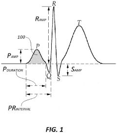
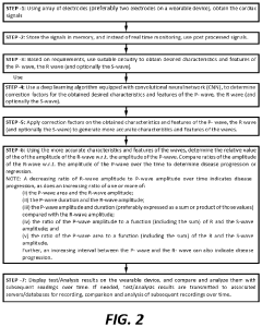
- A system and process for detecting cardiac amyloidosis using meticulously selected ECG parameters, which involves annotating a large database with cardiac amyloidosis diagnoses and using a machine learning model to identify informative ECG parameters such as P wave duration, P wave amplitude, R wave duration, and R wave amplitude.
Apparatus for Early Detection of Cardiac Amyloidosis
PatentPendingUS20220386928A1
Innovation
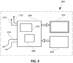
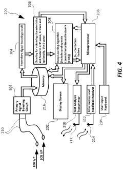
- A wearable device that determines the relative ratios of P-wave and R-wave features over time, such as P-wave area to R-wave amplitude, P-wave duration to R-wave amplitude, and P-wave amplitude to R-wave amplitude, allowing for early detection and surveillance of cardiac amyloidosis without requiring absolute R-wave measurements, using a microprocessor to record and compare these ratios.
Regulatory Framework for Cardiac Diagnostic Tools
The regulatory framework for cardiac diagnostic tools plays a crucial role in ensuring the safety, efficacy, and quality of devices used in diagnosing cardiac conditions, including P wave examination in cardiac amyloidosis patients. In the United States, the Food and Drug Administration (FDA) is the primary regulatory body overseeing medical devices, including those used for cardiac diagnostics.
The FDA classifies medical devices into three categories based on their risk level and intended use. Cardiac diagnostic tools typically fall under Class II or Class III, requiring more stringent regulatory oversight. For P wave examination devices, manufacturers must adhere to the 510(k) premarket notification process or the more rigorous Premarket Approval (PMA) pathway, depending on the device's classification.
Key regulatory requirements for cardiac diagnostic tools include demonstrating safety and effectiveness through clinical trials, implementing quality management systems, and adhering to Good Manufacturing Practices (GMP). Manufacturers must also comply with labeling requirements, post-market surveillance, and reporting of adverse events.
In the European Union, the regulatory landscape for cardiac diagnostic tools is governed by the Medical Device Regulation (MDR). The MDR, which replaced the previous Medical Device Directive (MDD), introduced more stringent requirements for clinical evidence, post-market surveillance, and traceability of medical devices.
For P wave examination devices used in cardiac amyloidosis patients, manufacturers must obtain CE marking to market their products in the EU. This process involves demonstrating compliance with the MDR's essential requirements, including clinical evaluation, risk management, and technical documentation.
Globally, regulatory bodies are increasingly focusing on the integration of software and artificial intelligence in cardiac diagnostic tools. This shift has led to the development of new guidelines and regulations specifically addressing software as a medical device (SaMD) and AI/ML-based medical devices.
As the field of cardiac diagnostics continues to evolve, regulatory frameworks are adapting to keep pace with technological advancements. This includes the development of new guidance documents, international harmonization efforts, and the implementation of unique device identification (UDI) systems to enhance traceability and patient safety.
Ethical Implications in Amyloidosis Diagnosis and Management
The ethical implications of P wave examination in cardiac amyloidosis patients extend beyond the technical aspects of diagnosis and management. As this advanced cardiac imaging technique becomes more prevalent, healthcare providers and researchers must navigate complex ethical considerations to ensure patient welfare and maintain the integrity of medical practice.
One primary ethical concern is the potential for overdiagnosis and unnecessary interventions. P wave examination may detect subtle cardiac abnormalities that might not progress to clinically significant disease. This raises questions about the appropriate threshold for intervention and the potential psychological impact on patients who receive a diagnosis of cardiac amyloidosis, even if it may not affect their quality of life in the near term.
Informed consent is another critical ethical issue in the context of P wave examination. Patients must be fully aware of the implications of undergoing this diagnostic procedure, including the possibility of incidental findings and the potential impact on their insurability or employment prospects. Healthcare providers have a responsibility to communicate clearly about the benefits, risks, and limitations of the examination.
Privacy and data protection concerns also come to the forefront with the increasing use of advanced imaging techniques. The detailed cardiac data obtained through P wave examination must be securely stored and protected from unauthorized access. Additionally, the use of this data for research purposes raises questions about patient consent and the appropriate balance between individual privacy and the potential for scientific advancement.
The equitable distribution of P wave examination technology is an important ethical consideration. As this diagnostic tool becomes more sophisticated and potentially more expensive, there is a risk of exacerbating healthcare disparities. Ensuring fair access to this technology across different socioeconomic groups and geographic regions is crucial to maintain ethical standards in healthcare delivery.
Furthermore, the interpretation of P wave examination results requires specialized expertise. There is an ethical obligation for healthcare systems to ensure that professionals interpreting these results are adequately trained and maintain their competence over time. Misinterpretation of results could lead to inappropriate treatment decisions and harm to patients.
Lastly, the development and implementation of P wave examination techniques must be guided by robust ethical frameworks in research. This includes ensuring that clinical trials are conducted with proper oversight, that results are reported transparently, and that negative findings are not suppressed. The scientific community has a responsibility to maintain high ethical standards in the pursuit of improved diagnostic and treatment options for cardiac amyloidosis patients.
Unlock deeper insights with Patsnap Eureka Quick Research — get a full tech report to explore trends and direct your research. Try now!
Generate Your Research Report Instantly with AI Agent
Supercharge your innovation with Patsnap Eureka AI Agent Platform!