Use of P wave in identifying heart valve disorders
AUG 19, 20259 MIN READ
Generate Your Research Report Instantly with AI Agent
Patsnap Eureka helps you evaluate technical feasibility & market potential.
P Wave Analysis Background
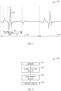
The P wave in electrocardiography (ECG) represents atrial depolarization and provides valuable information about the electrical activity of the heart's upper chambers. Historically, P wave analysis has been primarily used to diagnose atrial arrhythmias and conduction abnormalities. However, recent advancements in signal processing and machine learning techniques have expanded its potential applications, including the identification of heart valve disorders.
The concept of using P wave characteristics to detect valvular heart disease emerged from the understanding that structural and functional changes in heart valves can affect atrial electrical activity. As valvular disorders progress, they often lead to atrial remodeling, which in turn alters the morphology and timing of the P wave. This relationship between valve function and P wave characteristics forms the foundation for using P wave analysis as a diagnostic tool for heart valve disorders.
Early studies in this field focused on manual measurements of P wave duration, amplitude, and morphology. These parameters were found to correlate with specific valvular conditions, such as mitral stenosis and aortic regurgitation. However, the sensitivity and specificity of these manual measurements were limited, prompting researchers to explore more advanced analytical methods.
The advent of digital ECG systems and computerized signal processing techniques marked a significant turning point in P wave analysis. These technologies enabled more precise and reproducible measurements of P wave parameters, including P wave dispersion, P wave terminal force, and P wave area. Additionally, the development of advanced algorithms allowed for the extraction of subtle features that were previously undetectable through visual inspection alone.
Machine learning and artificial intelligence have further revolutionized P wave analysis in recent years. These technologies can identify complex patterns and relationships within P wave data that may not be apparent to human observers. By training algorithms on large datasets of ECGs from patients with known valvular disorders, researchers have developed models capable of detecting and classifying various heart valve abnormalities with increasing accuracy.
The potential of P wave analysis in identifying heart valve disorders extends beyond traditional ECG interpretation. Integration with other diagnostic modalities, such as echocardiography and cardiac MRI, has shown promise in enhancing the overall diagnostic accuracy and providing a more comprehensive assessment of valvular function. This multimodal approach leverages the strengths of each technique to overcome individual limitations and provide a more robust evaluation of cardiac health.
As research in this field continues to evolve, there is growing interest in the use of P wave analysis for early detection and monitoring of valvular heart disease. The non-invasive nature of ECG recordings, combined with the potential for automated analysis, makes this approach particularly attractive for large-scale screening programs and long-term patient monitoring. However, challenges remain in standardizing analysis methods and validating their clinical utility across diverse patient populations.
Clinical Need Assessment
The use of P wave analysis in identifying heart valve disorders addresses a critical need in cardiovascular diagnostics. Heart valve disorders affect millions of people worldwide, with an estimated prevalence of 2.5% in the general population. These disorders can lead to serious complications if left undiagnosed and untreated, including heart failure, stroke, and sudden cardiac death. Early detection and accurate diagnosis are crucial for effective management and improved patient outcomes.
Current diagnostic methods for heart valve disorders primarily rely on echocardiography, which, while effective, can be costly and time-consuming. Additionally, echocardiography requires specialized equipment and trained personnel, limiting its accessibility in resource-constrained settings. This creates a significant gap in the early detection and monitoring of heart valve disorders, particularly in primary care and rural healthcare facilities.
The P wave, representing atrial depolarization in the electrocardiogram (ECG), has shown promise as a potential biomarker for heart valve disorders. ECG is a widely available, non-invasive, and cost-effective diagnostic tool that can be easily performed in various healthcare settings. Leveraging P wave analysis for identifying heart valve disorders could significantly enhance the early detection capabilities of healthcare providers, especially in primary care settings.
There is a growing clinical need for more accessible and efficient screening methods for heart valve disorders. P wave analysis could potentially serve as a valuable initial screening tool, helping to identify patients who require further evaluation with more advanced diagnostic techniques. This approach could lead to earlier interventions, reduced healthcare costs, and improved patient outcomes.
Furthermore, the aging population in many countries is expected to increase the prevalence of heart valve disorders in the coming years. This demographic shift underscores the importance of developing innovative diagnostic approaches that can be easily integrated into routine clinical practice. P wave analysis offers a promising solution to meet this growing demand for efficient and scalable screening methods.
The potential applications of P wave analysis extend beyond initial screening. It could also be valuable in monitoring disease progression and assessing the effectiveness of treatments over time. This longitudinal monitoring capability could significantly enhance the management of heart valve disorders, allowing for more personalized and timely interventions.
In conclusion, the clinical need for improved methods of identifying heart valve disorders is substantial and growing. The use of P wave analysis presents an opportunity to address this need by providing a widely accessible, cost-effective, and potentially powerful diagnostic tool. Further research and development in this area could lead to significant advancements in cardiovascular care, benefiting patients and healthcare systems alike.
Current Challenges
The use of P waves in identifying heart valve disorders faces several significant challenges that hinder its widespread adoption and effectiveness in clinical practice. One of the primary obstacles is the complexity of accurately detecting and interpreting P waves in electrocardiograms (ECGs). P waves are often small in amplitude and can be obscured by noise or other cardiac electrical activities, making their precise identification challenging.
The variability in P wave morphology among different individuals and even within the same patient over time poses another substantial challenge. This variability can lead to difficulties in establishing standardized criteria for diagnosing specific heart valve disorders based solely on P wave characteristics. Consequently, the sensitivity and specificity of P wave analysis for valve disorder detection may be compromised.
Another critical challenge lies in the limited correlation between P wave abnormalities and specific valve disorders. While certain P wave changes can indicate atrial enlargement or conduction abnormalities, which may be associated with valve problems, the direct link between P wave characteristics and valve dysfunction is not always clear-cut. This ambiguity can result in potential misdiagnoses or overlooked valve issues if relying exclusively on P wave analysis.
The influence of other cardiac and non-cardiac factors on P wave morphology further complicates its use in valve disorder identification. Conditions such as hypertension, pulmonary diseases, and electrolyte imbalances can affect P wave characteristics, potentially leading to false positives or negatives in valve disorder diagnosis. Distinguishing between these confounding factors and true valve-related P wave changes remains a significant challenge.
Technical limitations in ECG recording and analysis systems also present obstacles. Many current ECG machines and algorithms are not optimized for detailed P wave analysis, focusing instead on more prominent features like QRS complexes. This limitation can result in inadequate resolution or inaccurate representation of P waves, hampering their utility in valve disorder identification.
The need for specialized training and expertise in P wave interpretation for valve disorder diagnosis is another hurdle. Many healthcare providers may not have the necessary skills or experience to accurately analyze P wave subtleties in the context of valve disorders, potentially leading to missed diagnoses or inappropriate patient management.
Lastly, the lack of large-scale, longitudinal studies validating the effectiveness of P wave analysis in identifying specific valve disorders across diverse patient populations remains a significant challenge. Without robust clinical evidence, the widespread adoption of this technique in routine cardiac care may be limited, despite its potential benefits.
Existing P Wave Solutions
01 P wave analysis for heart valve disorder detection
P wave analysis techniques are used to detect and diagnose heart valve disorders. These methods involve examining the characteristics of the P wave in electrocardiograms (ECGs) to identify abnormalities associated with various valve conditions. Advanced signal processing and machine learning algorithms may be employed to enhance the accuracy of detection and classification of heart valve disorders based on P wave morphology.- P wave analysis for heart valve disorder detection: P wave analysis techniques are used to detect and diagnose heart valve disorders. These methods involve examining the characteristics of the P wave in electrocardiograms (ECGs) to identify abnormalities associated with various valve conditions. Advanced signal processing and machine learning algorithms may be employed to enhance the accuracy of detection and classification of heart valve disorders based on P wave morphology.
- Implantable devices for heart valve monitoring: Implantable medical devices are developed to continuously monitor heart valve function and detect disorders. These devices may include sensors to measure various parameters related to valve performance, such as blood flow, pressure, and electrical activity. The collected data can be analyzed to identify potential valve disorders and alert healthcare providers for timely intervention.
- Non-invasive imaging techniques for valve assessment: Advanced non-invasive imaging techniques are utilized to assess heart valve function and detect disorders. These may include echocardiography, cardiac MRI, and CT scans. Image processing algorithms and artificial intelligence are employed to analyze the imaging data and identify subtle changes in valve structure and function that may indicate the presence of disorders.
- Wearable devices for continuous heart monitoring: Wearable devices are developed to provide continuous monitoring of heart activity, including P wave analysis for valve disorder detection. These devices may incorporate ECG sensors and advanced algorithms to analyze heart rhythms and detect abnormalities associated with valve disorders. The collected data can be transmitted to healthcare providers for remote monitoring and early intervention.
- Artificial intelligence for valve disorder prediction: Artificial intelligence and machine learning algorithms are developed to predict the onset of heart valve disorders based on various physiological parameters, including P wave characteristics. These systems analyze large datasets of patient information to identify patterns and risk factors associated with valve disorders, enabling early detection and personalized treatment strategies.
02 Implantable devices for heart valve monitoring
Implantable medical devices are developed to monitor heart valve function and detect disorders. These devices may include sensors to measure various parameters related to valve performance, such as blood flow, pressure, and electrical activity. The collected data can be analyzed to identify potential valve disorders and provide early warning signs for medical intervention.Expand Specific Solutions03 Non-invasive imaging techniques for valve assessment
Advanced non-invasive imaging techniques are utilized to assess heart valve function and diagnose disorders. These may include echocardiography, cardiac MRI, and CT scans. Image processing algorithms and artificial intelligence are employed to analyze the imaging data and detect subtle changes in valve structure and function that may indicate the presence of disorders.Expand Specific Solutions04 Wearable devices for continuous heart monitoring
Wearable devices are developed to provide continuous monitoring of heart activity, including P wave analysis for valve disorder detection. These devices may incorporate ECG sensors, accelerometers, and other biosensors to collect comprehensive data on heart function. The collected data is analyzed in real-time or periodically to identify potential valve disorders and alert users or healthcare providers.Expand Specific Solutions05 Artificial heart valves with integrated monitoring capabilities
Advanced artificial heart valves are designed with integrated sensors and monitoring capabilities. These smart valves can provide real-time data on their own function and the surrounding cardiac environment. The collected information can be used to detect potential complications or disorders related to the valve or the heart's overall performance, enabling timely interventions and adjustments.Expand Specific Solutions
Key Industry Players
The use of P waves in identifying heart valve disorders represents an emerging field in cardiac diagnostics, currently in its early development stage. The market size is relatively small but growing, driven by increasing prevalence of heart valve diseases and demand for non-invasive diagnostic tools. Technologically, the field is still evolving, with varying levels of maturity among key players. Companies like Medtronic, Boston Scientific, and Koninklijke Philips are leveraging their established presence in cardiac care to advance P wave analysis techniques. Smaller, specialized firms such as Bardy Diagnostics and Biosense Webster are focusing on innovative P wave monitoring devices. Chinese companies like Helowin Medical and HeartVoice are also making strides in AI-driven ECG analysis, potentially accelerating the technology's development and adoption.
Medtronic, Inc.
Technical Solution: Medtronic has developed advanced P-wave sensing algorithms for their cardiac devices to improve the detection of atrial arrhythmias and heart valve disorders. Their technology utilizes machine learning techniques to analyze P-wave morphology, duration, and amplitude changes[1]. This allows for early identification of potential valve issues, particularly mitral and tricuspid valve disorders. The system incorporates real-time P-wave analysis with other cardiac parameters to provide a comprehensive assessment of heart function[2]. Medtronic's implantable cardiac monitors and pacemakers are equipped with this technology, enabling continuous monitoring and early intervention for patients at risk of valve disorders.
Strengths: Highly accurate P-wave analysis, integration with existing cardiac devices, and continuous monitoring capabilities. Weaknesses: Requires invasive implantation of devices and may have limitations in detecting subtle valve abnormalities.
Boston Scientific Scimed, Inc.
Technical Solution: Boston Scientific has developed a novel P-wave analysis system for their cardiac rhythm management devices. Their technology focuses on analyzing P-wave variability and morphology changes to detect early signs of heart valve disorders, particularly those affecting the mitral and tricuspid valves[3]. The system employs advanced signal processing algorithms to filter out noise and isolate clear P-wave signals, even in challenging recording conditions[4]. Boston Scientific's approach also incorporates machine learning models trained on large datasets of P-wave patterns associated with various valve disorders. This allows for improved accuracy in distinguishing between normal variations and pathological changes indicative of valve problems[5].
Strengths: High sensitivity in detecting subtle P-wave changes, robust noise reduction capabilities, and integration with existing cardiac devices. Weaknesses: May require frequent algorithm updates and could be affected by other cardiac conditions that alter P-wave morphology.
Innovative P Wave Techniques
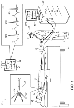
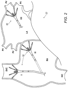
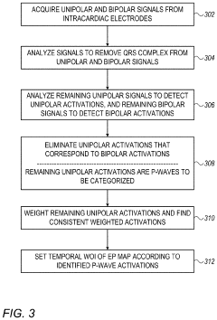
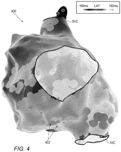
P-wave detection using intracardiac electrodes
PatentActiveUS20230284960A1
Innovation
- The use of intracardiac signals acquired by a mapping catheter, particularly from the inferior vena cava or superior vena cava, to detect far-field P-waves by removing dominant QRS complex signals and analyzing unipolar signals for P-wave activations, with a processor-based algorithm to eliminate near-field activations and weight signals for consistency, allowing for accurate P-wave identification and automatic window of interest setting.
Identifying p wave oversensing
PatentInactiveUS20210076964A1
Innovation
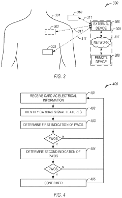
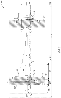
- A system and method that utilize a signal receiver circuit to receive cardiac electrical information, an assessment circuit to identify cardiac signal features, and determine P wave oversensing through a pattern analysis followed by a morphology confirmation, optimizing resource usage while maintaining sensitivity and specificity.
Regulatory Considerations
The use of P wave analysis in identifying heart valve disorders is subject to various regulatory considerations that must be carefully addressed. In the United States, the Food and Drug Administration (FDA) plays a crucial role in overseeing medical devices and diagnostic tools. Any technology utilizing P wave analysis for heart valve disorder detection would likely fall under Class II medical devices, requiring a 510(k) premarket notification submission to demonstrate substantial equivalence to a legally marketed predicate device.
Regulatory bodies emphasize the importance of clinical validation studies to establish the accuracy and reliability of P wave analysis in detecting heart valve disorders. These studies must adhere to Good Clinical Practice (GCP) guidelines and may require Institutional Review Board (IRB) approval. The FDA's guidance on Software as a Medical Device (SaMD) is particularly relevant, as P wave analysis often involves complex algorithms and software systems.
Data privacy and security regulations, such as the Health Insurance Portability and Accountability Act (HIPAA) in the United States and the General Data Protection Regulation (GDPR) in the European Union, must be strictly followed. These regulations govern the collection, storage, and transmission of patient data, including electrocardiogram (ECG) recordings used for P wave analysis.
International standards, such as ISO 13485 for quality management systems in medical devices and IEC 62304 for medical device software lifecycle processes, are essential for ensuring compliance and facilitating global market access. Manufacturers must also consider the International Medical Device Regulators Forum (IMDRF) guidelines, which aim to harmonize regulatory approaches across different countries.
Post-market surveillance is another critical regulatory aspect. Manufacturers must implement systems to monitor the performance and safety of their P wave analysis technology in real-world settings. This includes reporting adverse events and maintaining vigilance for potential issues that may arise during widespread use.
As the field of cardiac diagnostics evolves, regulatory bodies may update their guidelines to address emerging technologies. Companies developing P wave analysis tools for heart valve disorder identification must stay informed about these changes and adapt their regulatory strategies accordingly. Engaging with regulatory experts and maintaining open communication with relevant authorities can help navigate the complex landscape of medical device regulations.
AI Integration Prospects
The integration of artificial intelligence (AI) in the use of P waves for identifying heart valve disorders presents a promising frontier in cardiovascular diagnostics. Machine learning algorithms can be trained on large datasets of P wave morphologies to detect subtle patterns indicative of specific valve abnormalities. This approach has the potential to significantly enhance the accuracy and speed of diagnosis, particularly in cases where traditional methods may be inconclusive.
Deep learning models, such as convolutional neural networks (CNNs), can be applied to analyze the complex waveforms of P waves, extracting features that may not be apparent to the human eye. These models can be trained to recognize minute variations in P wave duration, amplitude, and morphology that correlate with different types of heart valve disorders. The ability of AI to process and interpret vast amounts of data quickly could lead to more efficient screening processes and earlier detection of valve problems.
AI-powered systems could also be developed to integrate P wave analysis with other cardiac parameters and patient data, providing a more comprehensive assessment of heart valve function. This holistic approach could improve risk stratification and guide personalized treatment strategies. Furthermore, AI algorithms could be designed to continuously learn and adapt as new data becomes available, potentially improving diagnostic accuracy over time.
The implementation of AI in this field could extend beyond traditional clinical settings. Wearable devices equipped with ECG sensors and AI capabilities could enable continuous monitoring of P waves in real-time, alerting patients and healthcare providers to potential valve issues before they become symptomatic. This proactive approach could revolutionize preventive cardiology and reduce the incidence of advanced heart valve disease.
However, the successful integration of AI in P wave analysis for heart valve disorders faces several challenges. Ensuring the reliability and interpretability of AI-generated diagnoses is crucial for clinical adoption. Rigorous validation studies and regulatory approvals will be necessary before widespread implementation. Additionally, addressing concerns about data privacy and security in AI-powered healthcare systems will be essential to gain patient trust and compliance with ethical standards.
Unlock deeper insights with Patsnap Eureka Quick Research — get a full tech report to explore trends and direct your research. Try now!
Generate Your Research Report Instantly with AI Agent
Supercharge your innovation with Patsnap Eureka AI Agent Platform!