
A Focusing Method of Camera Image in Digital Slice Scanner
A technology of digital slicing and focusing method, which is applied in the field of image processing, can solve problems such as multiple fields of view, image out of focus, and loss of focus, and achieve the effect of solving image out of focus and fast focusing speed
Patent Information
- Authority / Receiving Office
- CN · China
- Patent Type
- Patents(China)
- Current Assignee / Owner
- Publication Date
- 2021-08-24
Smart Images
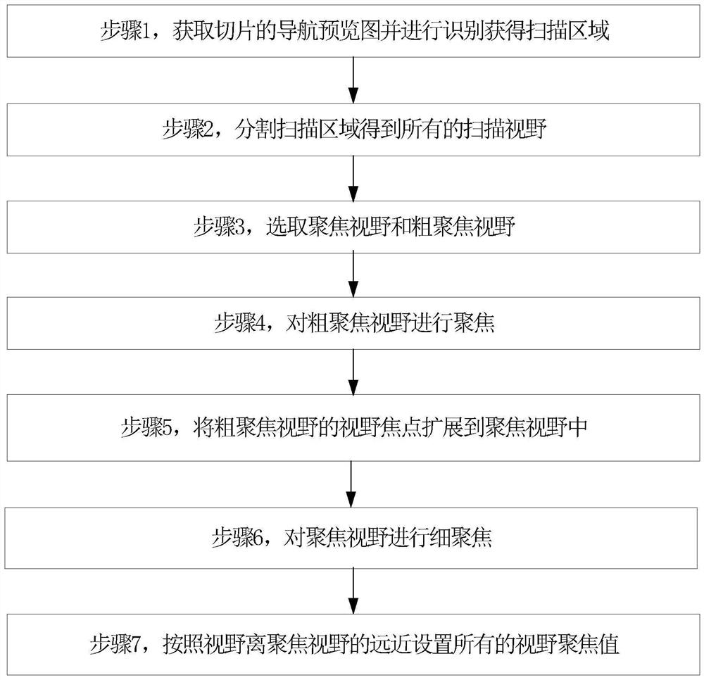
Figure 1 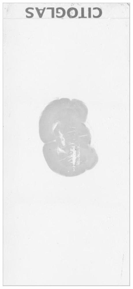
Figure 2 
Figure 3
Abstract
Description
technical field
[0001] The invention relates to a camera image focusing method in a digital slice scanner, belonging to the technical field of image processing. Background technique
[0002] Pathological examination is a pathological and morphological method used to examine pathological changes in body organs, tissues or cells. In order to explore the disease process of organs, tissues or cells, digital pathological slide scanners can be used to examine the pathological morphological examination of their lesions, and discuss the causes, pathogenesis, and development of lesions. process, and finally make a pathological diagnosis and pathological morphological examination.
[0003] The digital pathology slide scanner combines the micro-optical magnification system and computer technology to scan and collect images of traditional pathological glass slices one by one, and splicing and merging each pair of images to form a high-resolution, full-information digital pathology sli...
Examples
Embodiment Construction
[0046] Below in conjunction with accompanying drawing and embodiment the present invention will be further described:
[0047] In order to clearly illustrate the technical features of this solution, the present invention will be described in detail below through specific implementation modes and in conjunction with the accompanying drawings. The following disclosure provides many different embodiments or examples for implementing different structures of the present invention. To simplify the disclosure of the present invention, components and arrangements of specific examples are described below. Furthermore, the present invention may repeat reference numerals and / or letters in different instances. This repetition is for the purpose of simplicity and clarity and does not in itself indicate a relationship between the various embodiments and / or arrangements discussed. It should be noted that components illustrated in the figures are not necessarily drawn to scale. Description...