
Method for detecting human papillomavirus mrna
a human papillomavirus and detection method technology, applied in microbiological testing/measurement, biochemistry apparatus and processes, enzymes, etc., can solve the problems of low risk of these women, cell may soon develop some changes, and the possibility of regression of cell changes
Patent Information
- Authority / Receiving Office
- US · United States
- Patent Type
- Applications(United States)
- Current Assignee / Owner
- Publication Date
- 2005-06-02
- Estimated Expiration
- Not applicable · inactive patent
Smart Images
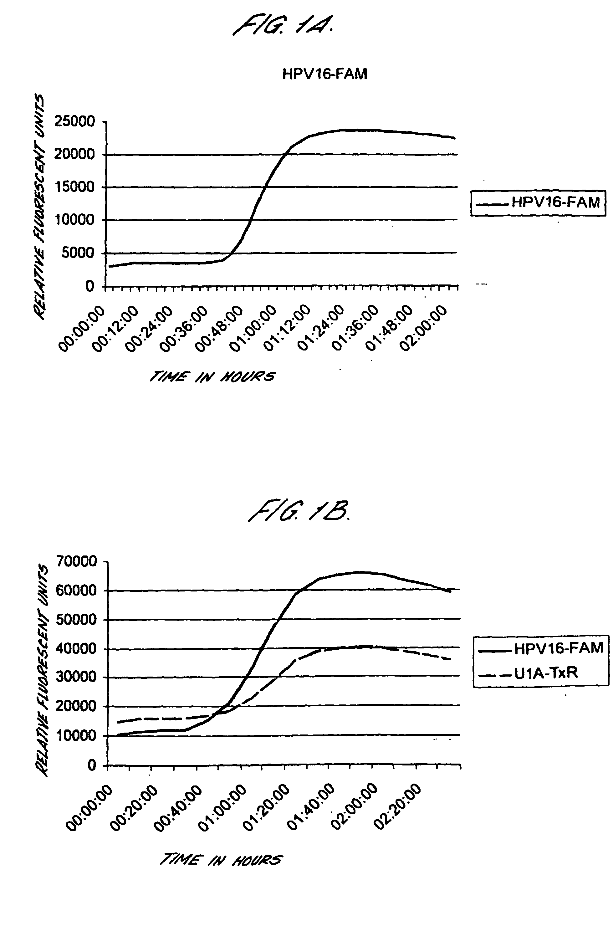
Figure 1 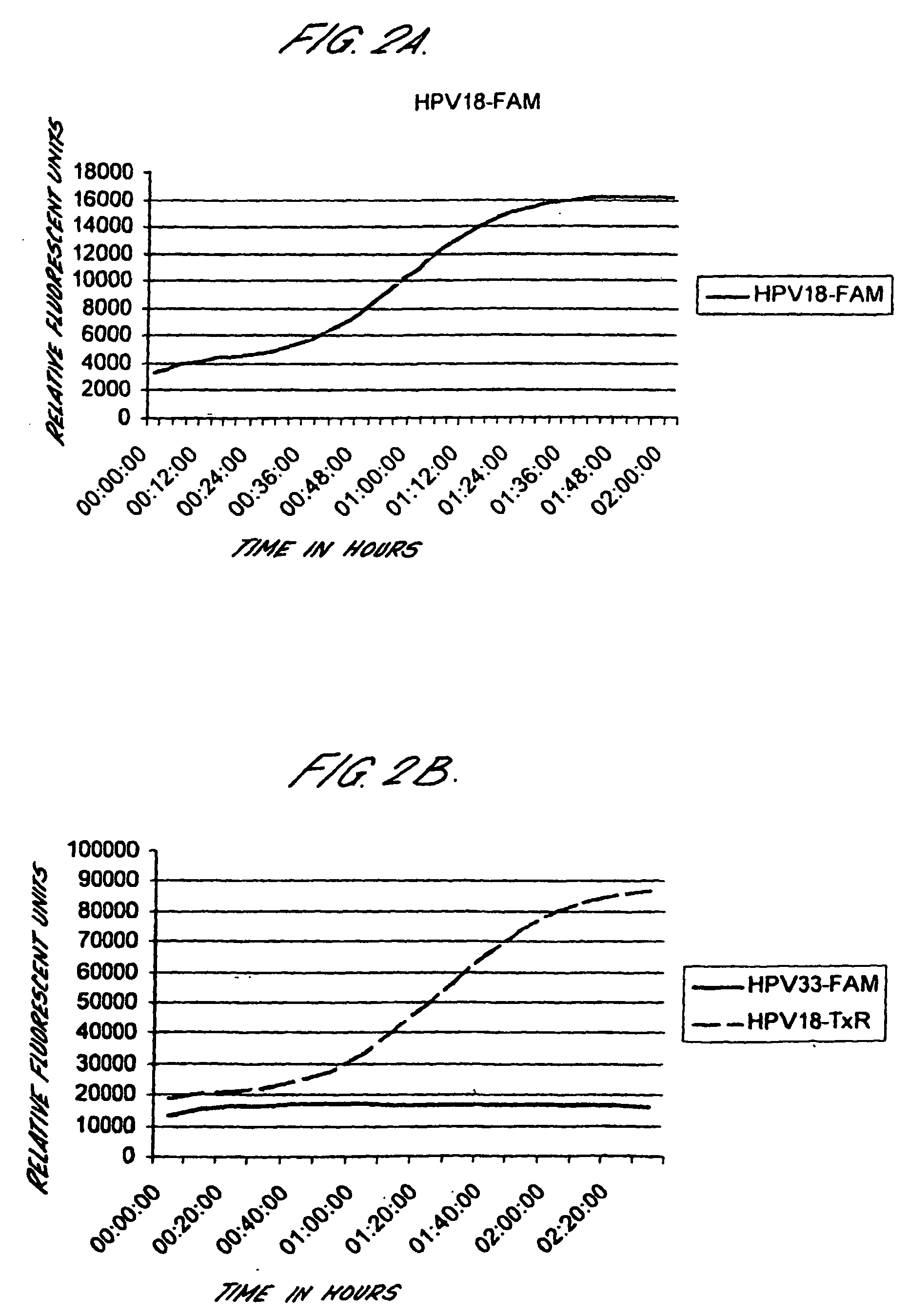
Figure 2 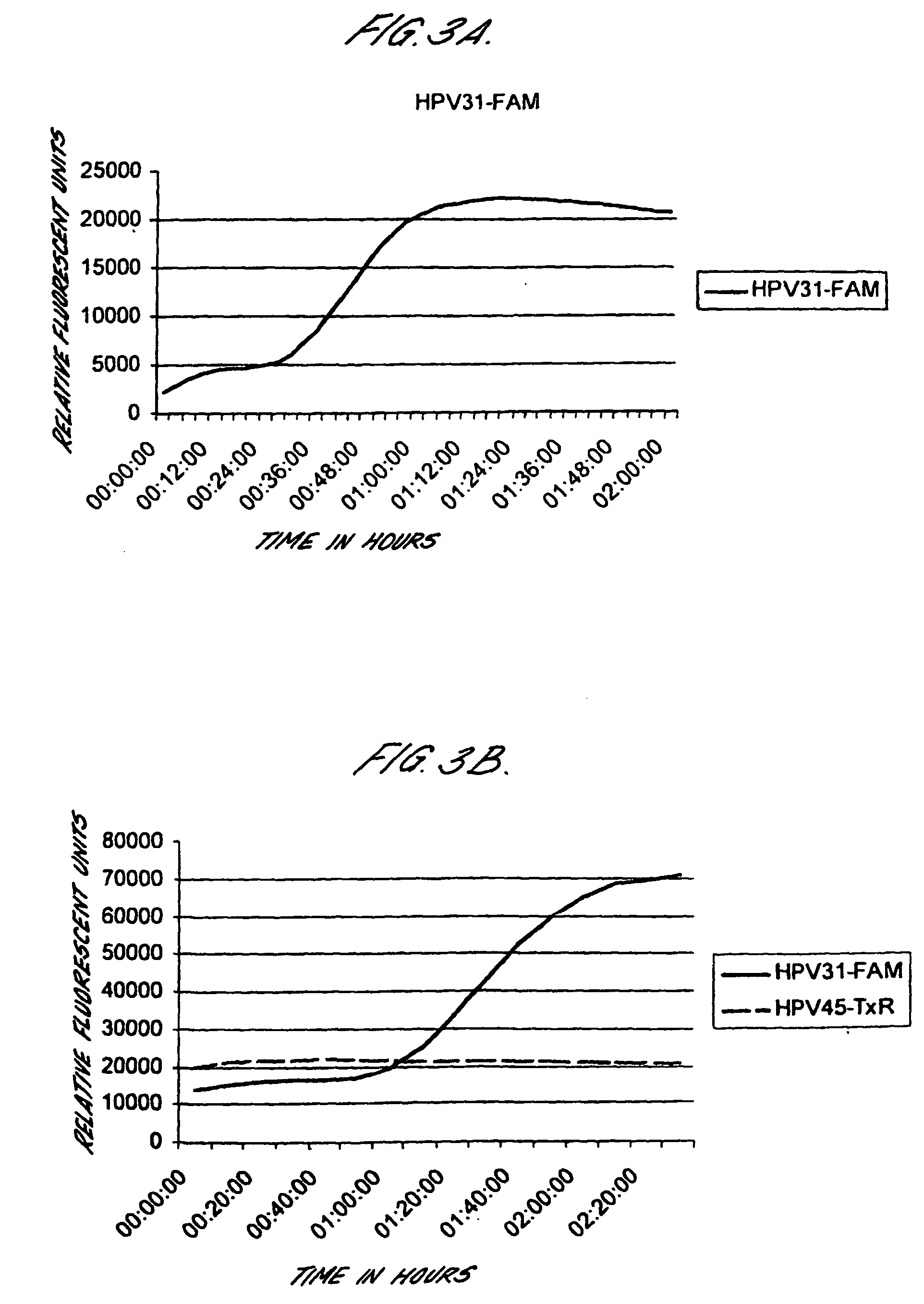
Figure 3
Abstract
Description
FIELD OF THE INVENTION
[0001] The present invention relates to in vitro methods of screening human subjects in order to assess their risk of developing cervical carcinoma. BACKGROUND TO THE INVENTION
[0002] Cervical carcinoma is one of the most common malignant diseases world-wide and is one of the leading causes of morbidity and mortality among women (Parkin D M, Pisani P, Ferlay J (1993) Int J Cancer 54: 594-606;Pisani P, Parkin D M, Ferlay J (1993) Int J Cancer 55: 891-903). 15,700 new cases of invasive cervical cancer were predicted in the United States in 1996, and the annual world-wide incidence is estimated to be 450,000 by the World Health Organization (1990). The annual incidence rate differs in different parts of the world, ranging from 7.6 per 100,000 in western Asia to 46.8 per 100,000 in southern Africa (Parkin et al., 1993 ibid).
[0003] The current conception of cervical carcinoma is that it is a multistage disease, often developing over a period of 10-25 years. Invasi...
Examples
example 2
Sensitivity of Real-Time NASBA on Control Cell Lines
[0191] Cervical cancer cell lines, CaSki, SiHa and HeLa were diluted in lysis buffer either before automated extraction of nucleic acids using the Boom's extraction method from Organon Teknika / bioMerieux (parallels 1 and 3), or after nucleic acid extraction (parallel 2). Real-time NASBA was performed using molecular beacons probes labelled with Texas red (16, L1 and 18) or FAM (U1A, 33 and 31) following the protocol described above.
TABLE 9PrimerCaSkiCaSkiHeLasets and161616333333183118311831probesE6U1E6U1E6U1L1E6L1E6L1E6E6E6E6E6E6E6Parallels112233112233112233Number ofCells100 000+++++++−+−+−+−+−+− 10 000+++++++−+−+−+−+−+− 1 000+++++++−+−+−+−+−+− 100+++++++−+−+−+−+−+− 10++++++−−−−+−+−+−+− 1−−+−+−−−−−−−+−+−+− 10−1−−−−−−−−−−−−−−−−−−
[0192] Thus, it is possible to detect HPV E6 mRNA in less than 1 cell using real-time NASBA.
[0193] Real-time NASBA was tested both as a multiplex assay and as single reactions. The results from ...
example 3
Further Clinical Study in 190 Patients
Patients / Clinical Samples
[0223] Biopsies from 190 women admitted to Østfold central-hospital for treatment of CIN in the period 1999-2001. The mean age of the 190 women included in the study was 37.4 years (range 22-74 years). Biopsies were frozen in −80° C. immediately after collection.
Cytological Examination of Samples
[0224] The routine cytological reports were used to record cytological findings. No attempt was made to re-evaluate the slides. Each one of them indicated a CIN II-III condition, i.e. a high grade dysplasia or HSIL, which was the basis for hospital admittance, colposcopy and biopsy.
Histological Examination of Samples
[0225] A biopsy, here termed biopsy 1, was taken after a high-grade cytology report. If it confirmed a high-grade lesion (CIN II or III), the patient was again admitted to hospital, this time for colposcopically guided conization. Before the conization, but after local anesthesia was applied, a second biopsy ...
example 4
HPV Detected by PreTect HPV-Proofer and PCR Compared to Cytology and Histology:
[0235] Normal and ASCUS samples (including borderline smears) were determined by cytology. All samples were tested with consensus PCR and PreTect HPV-Proofer but only the consensus positive samples were typed by PCR. The CIN 3 and cancer samples were determined by histology and all the samples were tested with all three methods. The results are shown in FIG. 6. Concordance between real-time multiplex NASBA and PCR compared to cytology or histology is shown in Table 15 below.
TABLE 15Concordance between real-time multiplex NASBAand PCR compared to cytology or histologyCytology / HistologyConcordancea (Number)Concordanceb (Number)Normal98.2% (4043)42.8% (138)ASCUSc94.5% (55)78.6% (14)CIN 394.3% (53)93.2% (44)Cancer99.0% (196)98.8% (170)
Only samples positive by Gp5+ / 6+ PCR have been typed.
aIncluding PCR and real-time multiplex NASBA positive and negative samples.
bIncluding only PCR and / or real-time multip...