
Quantification of microsphere suspension hybridization and uses thereof
a technology of microsphere suspension and hybridization, which is applied in the field of detection of chromosomal abnormalities, and can solve the problems of reducing the analysis time and overall cost per sample tested, and reducing the amount of sample required per assay
Patent Information
- Authority / Receiving Office
- US · United States
- Patent Type
- Applications(United States)
- Current Assignee / Owner
- Publication Date
- 2009-05-28
- Estimated Expiration
- Not applicable · inactive patent
Smart Images
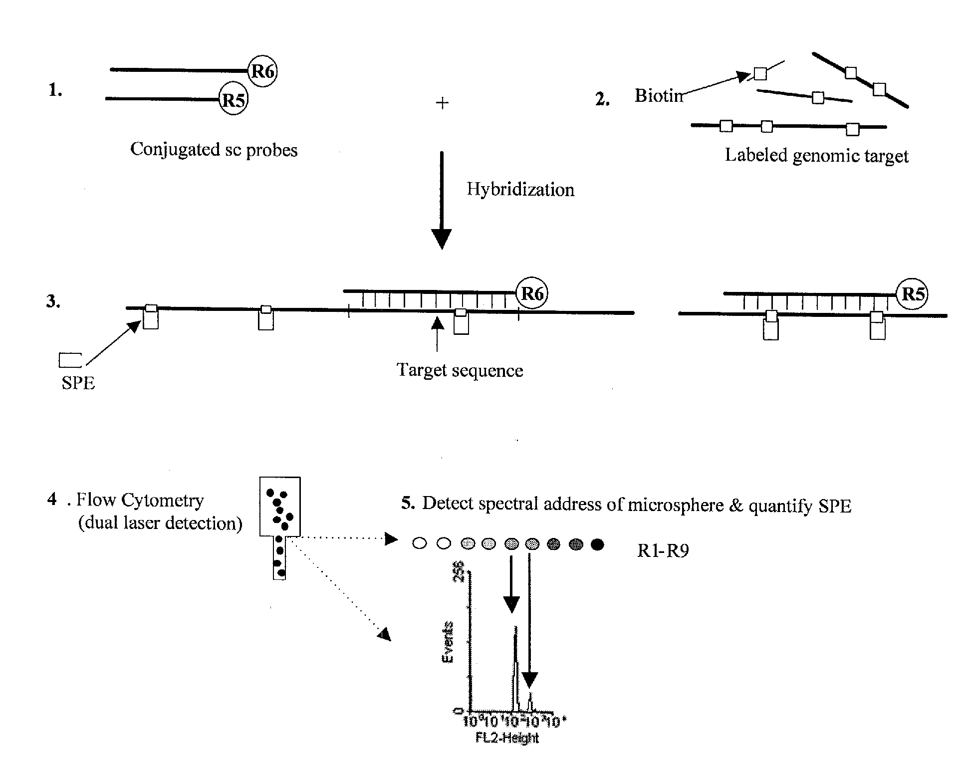
Figure 1 
Figure 2 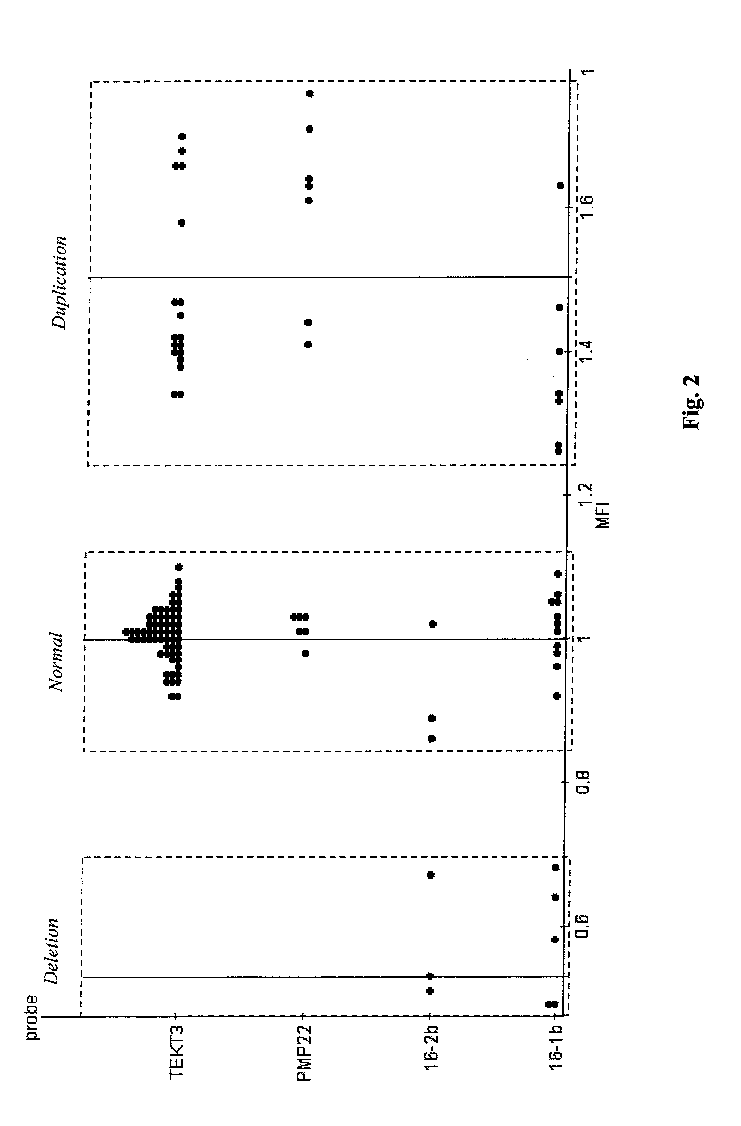
Figure 3
Abstract
Description
CROSS REFERENCE TO RELATED APPLICATION
[0001] This application claims the benefit of the prior filed, co-pending provisional application Ser. No. 60 / 708,734, filed Aug. 16, 2005, which is hereby incorporated by reference.SEQUENCE LISTING
[0002] A printed Sequence Listing, hereby incorporated by reference, accompanies this application, and has also been submitted with identical contents in the form of a computer-readable ASCII file on a floppy diskette.BACKGROUND OF THE INVENTION
[0003] 1. Field of the Invention
[0004] The present invention concerns materials and methods for the detection of chromosomal abnormalities using low copy nucleic acid hybridization probes. More particularly, the present invention concerns quantification of chromosomal abnormalities in nucleic acid sequences through hybridization of microsphere-conjugated, low copy nucleic acid probes to labeled target nucleic acid. Still more particularly, the present invention concerns conjugating a spectrally-encoded microsphere ...
Examples
example 1
References for Example 1
[0136]The following reference materials are hereby incorporated by reference.[0137]Armour J A L, Sismani C, Patsalis P C, Cross G. 2000. Measurement of locus copy number by hybridisation with amplifiable probes. Nucleic Acids Res. 28 (2):605-609.[0138]Bagwell C B, Baker D, Whetstone S, Munson M, Hitchcox S, Ault K A, Lovett E J. 1989. A simple and rapid method for determining the linearity of a flow cytometer amplification system. Cytometry 10 (6):689-94.[0139]Brown R D, Zarbo R J, Linden M D, Torres F X, Nakleh R E, Schultz D, Mackowiak P G. 1994. Two-color multiparametric method for flow cytometric DNA analysis. Standardization of spectral compensation. American Journal of Clinical Pathology 101 (5):630-637.[0140]Coder D M, Redelman D, Vogt R F. 1994. Computing the central location of immunofluorescence distributions: logarithmic data transformations are not always appropriate. Cytometry 18 (2):75-8.[0141]Dunbar S, Godbout R, Newkirk H, Hetzel J. 2003. Micr...
example 2
References for Example 2
[0161]The following reference materials are hereby incorporated by reference.[0162]Amos-Landgraf J M, Ji Y, Gottlieb W, Depinet T, Wandstrat A E, Cassidy S B, Driscoll D J, Rogan P K, Schwartz S, Nicholls R D (1999) Chromosome breakage in the Prader-Willi and Angelman syndromes involves recombination between large, transcribed repeats at proximal and distal breakpoints. Am J Hum Genet 65:370-386.[0163]Bejjani B A, Shaffer L G (2004) A cytogeneticist's perspective on genomic microarrays. Hum Reprod Update 10:221-226[0164]Bittel D C, Butler M G (2005) Prader-Willi syndrome: clinical genetics, cytogenetics and molecular biology. Expert Reviews in Molecular Medicine 7:1-20.[0165]Bittel D C, Kibiryeva N, Talebizadeh Z, Butler M G (2003) Microarray analysis of gene / transcript expression in Prader-Willi syndrome: deletion versus UPD. J Med Genet 40:568-574.[0166]Bittel D C, Kibiryeva N, Talebizadeh Z, Driscoll D J, Butler M G (2005) Microarray analysis of gene / trans...