
Method and Apparatus for Detecting Irregularities in Tissue Microarrays
a tissue microarray and irregularity detection technology, applied in the field of image processing and image analysis, can solve the problems of high time-consuming step of sequential multiplexing study, etc., and achieve the effect of facilitating efficient verification, tissue folding and loss
Patent Information
- Authority / Receiving Office
- US · United States
- Patent Type
- Applications(United States)
- Current Assignee / Owner
- Publication Date
- 2009-10-01
Smart Images
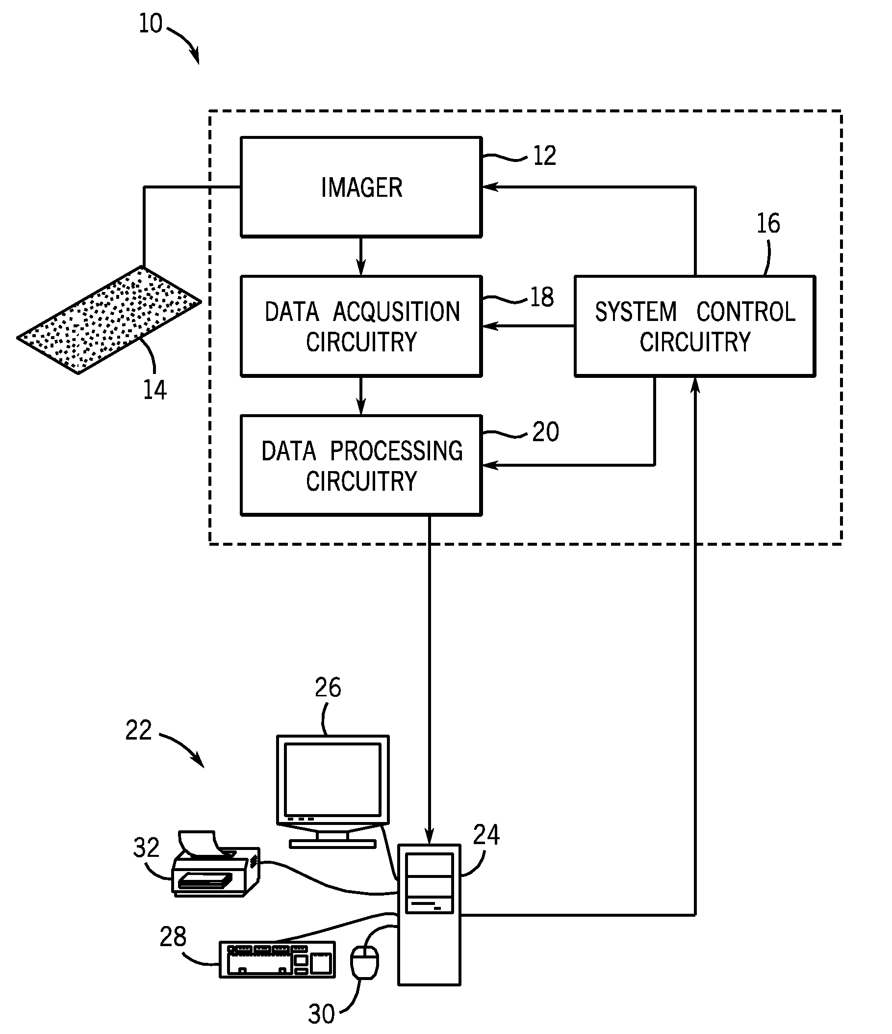
Figure 1 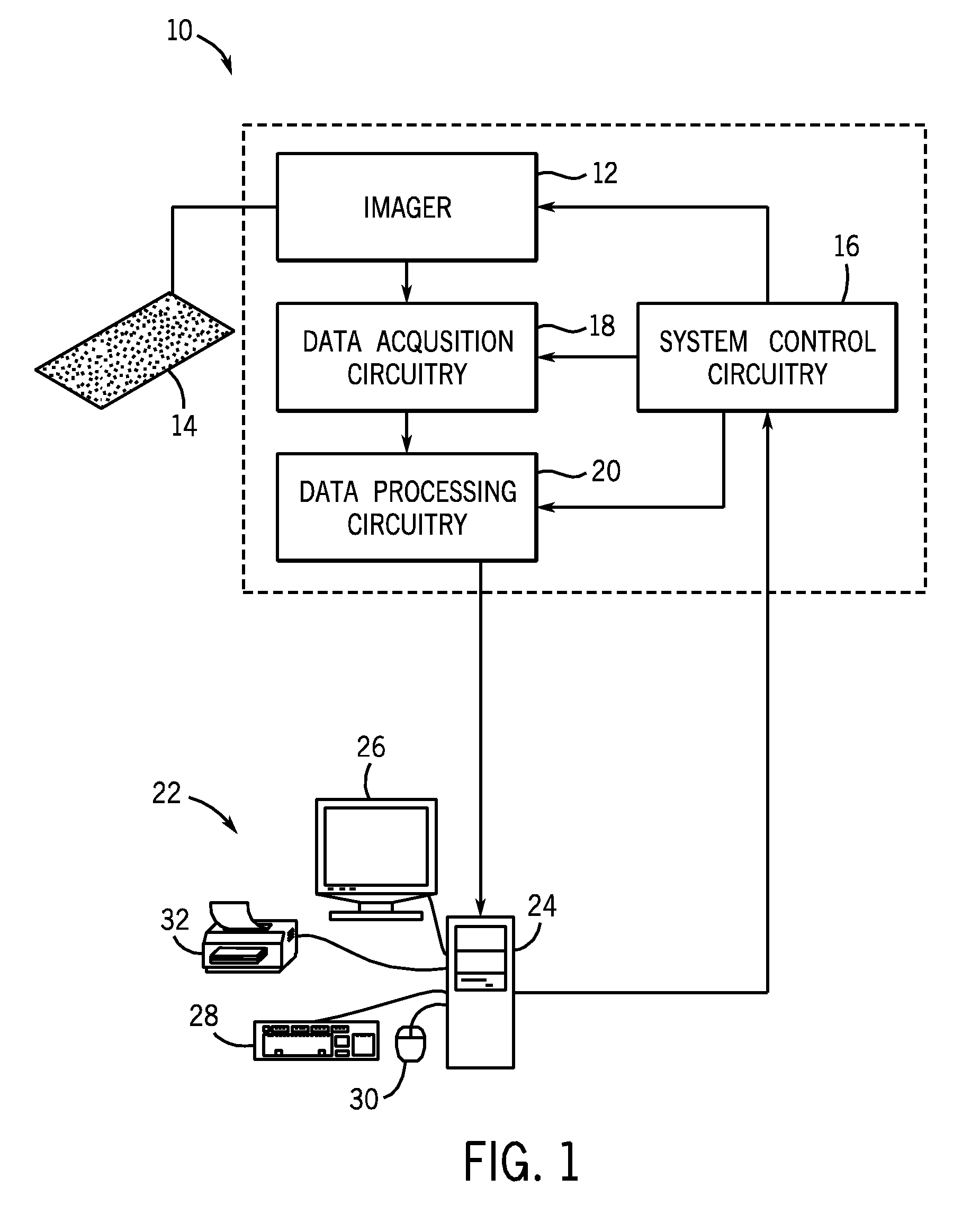
Figure 2 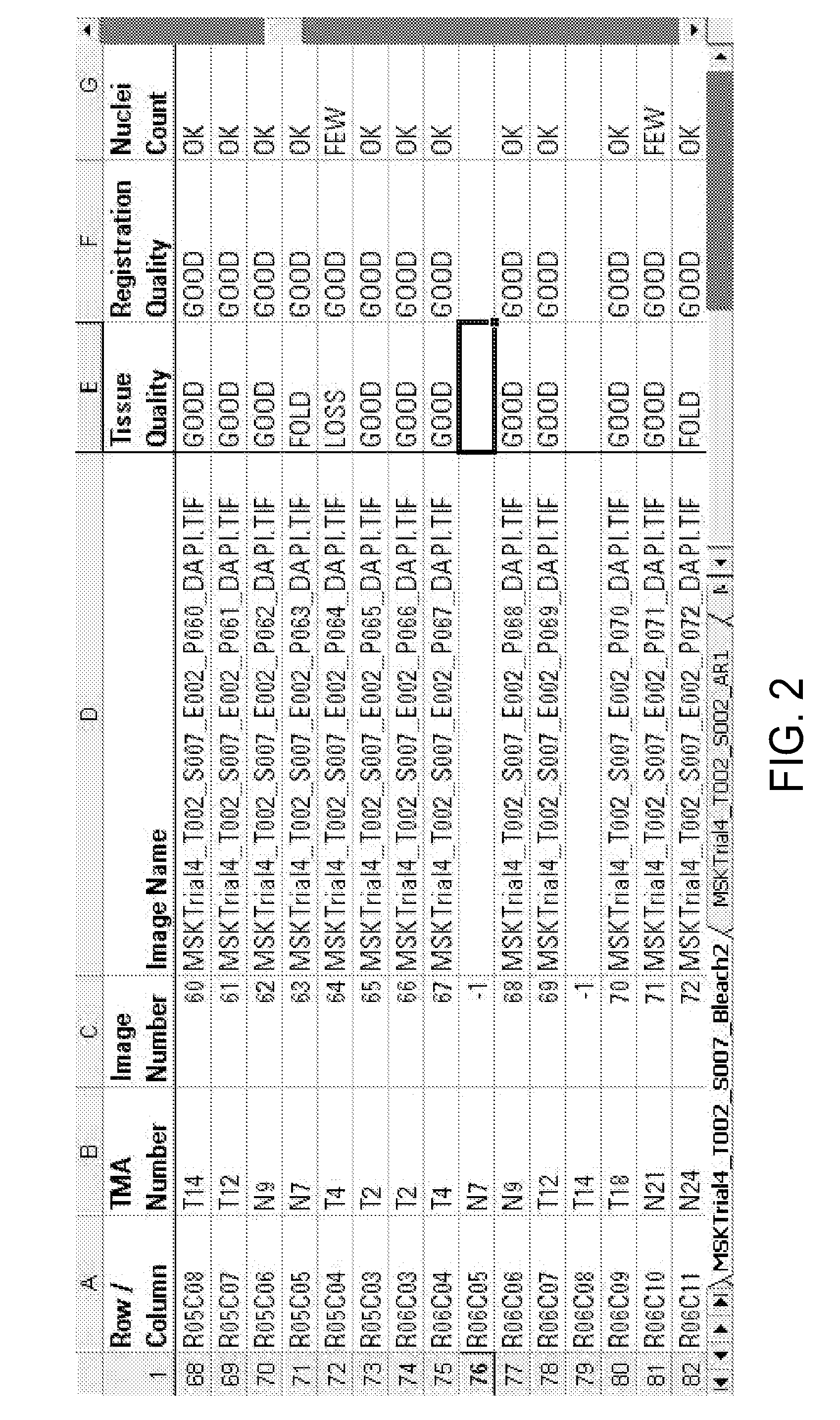
Figure 3
Abstract
Description
BACKGROUND
[0001] The invention relates generally to image processing and image analysis. More specifically, the present techniques relate to analysis of tissue microarrays.
[0002] Tissue microarrray (TMA) technology has become the standard in large-scale immunohistochemistry (IHC), fluorescent in situ hybridization (FISH), and mRNA in situ hybridization (RNA-ISH) studies for protein, DNA and RNA expression. To prepare the TMA slides, a tissue core is typically obtained from the patient tissue and inserted in a paraffin recipient block. The resulting recipient block typically has hundreds of tissue cores from multiple patients. This block may then be cut into sections with many different tissue spots corresponding to the tissue cores. The sections may be placed on glass slides for examination and imaging.
[0003] The development of TMA technology has generated interest in studies involving multiple biomarkers that may be performed on a single slide, e.g., sequential tissue multiplexing, te...
Examples
Embodiment Construction
[0029]The present techniques provide automated systems and methods for registering images of corresponding tissue spots in a TMA, detecting cases of registration failures, and / or re-initializing registration in the case of registration failure. The present techniques may reduce the incidence of individual validation of each tissue spot on a TMA. By providing a whole TMA image output, any tissue spot that failed to register may be annotated with a flag or other indicator. This output may allow an operator to scan an entire slide and quickly identify those tissue spots that may warrant additional validation and / or exclusion from further analysis.
[0030]The present techniques may use images of the tissue spots on a TMA and the relative x-y coordinates of each tissue spot as recorded by a microscope or other suitable image acquisition system. In certain embodiments, it is envisioned that the present techniques may be used in conjunction with previously acquired images, for example, digit...