
Mass spectrometric differentiation of tissue states
Patent Information
- Authority / Receiving Office
- US · United States
- Current Assignee / Owner
- Publication Date
- 2006-03-23
Smart Images
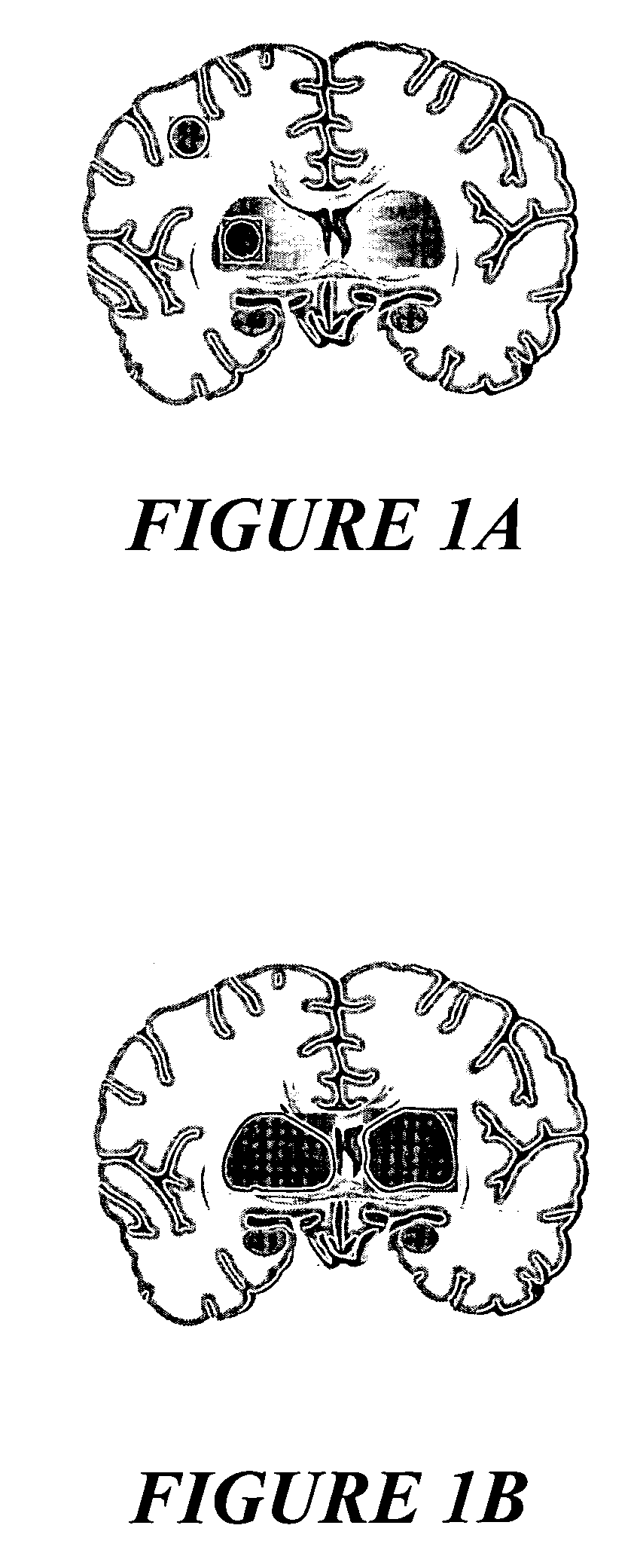
Figure 1
Abstract
Description
FIELD OF THE INVENTION
[0001] The invention relates to the determination and visualization of the spatial distribution of tissue states in histologic tissue sections on the basis of spatially resolved mass spectrometric signals. BACKGROUND OF THE INVENTION
[0002] The term “tissue state” here means the state of a small subarea of a tissue section with respect to a stress, a pathological change, an infection or other type of change compared with a normal state of this tissue. The tissue state must therefore be identifiable as a concentration pattern of substances which can be detected in this small subarea by a mass spectrometer. The substances can be peptides or proteins which are under- or overexpressed and hence form a pattern, or they can include positranslational modifications of proteins, their breakdown products (metabolites), or collections of other substances in the tissue.
[0003] Mass spectrometry with ionization of the samples by matrix-assisted laser desorption and ionizat...
Examples
Embodiment Construction
[0034] One preferred embodiment begins with the production of a microscopic tissue section, preferably from a deep-frozen piece of tissue, with a microtome. The microscopic tissue section is applied to a suitable support. This support can be a glass specimen slide, for example, whose surface is equipped with a transparent but conductive surface coating for subsequent use in the mass spectrometer. Other supports, for example metal supports or supports made of electrically conductive plastic, can also be used, however. The microscopic tissue section can then be stained in the usual way, although care has to be taken to use a stain which does not interfere with a subsequent mass spectrometric analysis of the tissue constituents. Fluorescence dyeing methods can also be used if they do not restrict the mass spectrometric analysis.
[0035] After this, a microscopic image is taken, with transmitted or reflected light, from the microscopic tissue section, and is later used to lay under the r...