Impact of P wave on atrial electrical activity
AUG 19, 20259 MIN READ
Generate Your Research Report Instantly with AI Agent
Patsnap Eureka helps you evaluate technical feasibility & market potential.
P Wave Background and Objectives
The P wave, a crucial component of the electrocardiogram (ECG), represents the electrical depolarization of the atria preceding atrial contraction. This electrical activity, originating from the sinoatrial node, plays a vital role in initiating the cardiac cycle and maintaining proper heart rhythm. Over the past century, our understanding of the P wave and its significance in atrial electrical activity has evolved significantly, shaping modern cardiology and electrophysiology.
The study of P waves dates back to the early 20th century when Willem Einthoven developed the string galvanometer, enabling the recording of electrical activity in the heart. Since then, technological advancements have allowed for more precise measurements and analysis of P wave morphology, duration, and amplitude. These improvements have led to a deeper understanding of atrial conduction patterns and their implications for cardiac health.
Recent years have witnessed a surge in research focusing on the impact of P waves on atrial electrical activity. This renewed interest stems from the growing prevalence of atrial fibrillation and other atrial arrhythmias, which pose significant health risks and economic burdens worldwide. As a result, there is an increasing need to comprehend the intricate relationship between P wave characteristics and atrial electrophysiology.
The primary objective of current research in this field is to elucidate the mechanisms by which P wave abnormalities influence atrial electrical activity and contribute to arrhythmogenesis. This includes investigating how variations in P wave morphology, duration, and amplitude correlate with specific atrial conduction disturbances and structural changes. Additionally, researchers aim to develop more accurate diagnostic tools and risk stratification methods based on P wave analysis.
Another critical goal is to explore the potential of P wave-based interventions in preventing and managing atrial arrhythmias. This involves studying how modulation of P wave characteristics through pharmacological or device-based therapies might alter atrial electrical properties and reduce the likelihood of arrhythmic events. Furthermore, there is a growing interest in utilizing advanced signal processing techniques and artificial intelligence to extract more information from P waves, potentially uncovering subtle markers of atrial dysfunction.
As technology continues to advance, the field is moving towards more comprehensive and integrated approaches to studying P waves and atrial electrical activity. This includes the development of high-resolution mapping techniques, computational modeling of atrial electrophysiology, and the integration of genetic and molecular data to provide a more holistic understanding of atrial function and dysfunction.
Clinical Significance of P Wave Analysis
The analysis of P waves in electrocardiograms (ECGs) holds significant clinical importance in the diagnosis and management of various cardiac conditions. P waves represent atrial depolarization and provide valuable insights into atrial electrical activity and conduction patterns. By carefully examining P wave morphology, duration, and amplitude, clinicians can detect and assess a wide range of atrial abnormalities and predict potential cardiac complications.
One of the primary clinical applications of P wave analysis is in the diagnosis of atrial enlargement. Left atrial enlargement is often associated with mitral valve disease, hypertension, and heart failure, while right atrial enlargement may indicate pulmonary hypertension or tricuspid valve disease. Characteristic changes in P wave morphology, such as increased amplitude or duration, can help identify these conditions early, allowing for timely intervention and management.
P wave analysis also plays a crucial role in the detection and evaluation of atrial arrhythmias. Abnormalities in P wave patterns can indicate various types of atrial conduction disturbances, including interatrial block, atrial fibrillation, and atrial flutter. Early identification of these arrhythmias through P wave analysis enables clinicians to initiate appropriate treatment strategies and prevent potential complications such as stroke or heart failure.
Furthermore, P wave analysis contributes to the assessment of atrioventricular (AV) conduction abnormalities. Changes in PR interval duration or the presence of dissociated P waves can indicate various degrees of AV block, which may require intervention such as pacemaker implantation. Additionally, P wave analysis can help differentiate between supraventricular and ventricular tachycardias, guiding appropriate management strategies.
In the context of risk stratification, P wave analysis has emerged as a valuable tool for predicting the likelihood of developing atrial fibrillation. Studies have shown that certain P wave characteristics, such as prolonged P wave duration or increased P wave dispersion, are associated with an increased risk of atrial fibrillation. This information allows clinicians to identify high-risk patients and implement preventive measures or closer monitoring.
P wave analysis also contributes to the evaluation of the effectiveness of antiarrhythmic therapies. Changes in P wave morphology and conduction patterns can provide insights into the response to treatment and guide necessary adjustments. Moreover, P wave analysis can aid in the assessment of successful cardioversion or ablation procedures for atrial arrhythmias.
In conclusion, the clinical significance of P wave analysis extends across various aspects of cardiac care, from diagnosis and risk stratification to treatment evaluation and long-term management. As our understanding of atrial electrical activity continues to evolve, P wave analysis remains a fundamental tool in clinical cardiology, providing valuable information for improved patient outcomes and personalized treatment strategies.
Current Challenges in P Wave Detection
P wave detection in electrocardiograms (ECGs) plays a crucial role in understanding atrial electrical activity and diagnosing various cardiac conditions. However, several challenges persist in accurately identifying and analyzing P waves, hindering the comprehensive assessment of atrial function.
One of the primary challenges in P wave detection is the low amplitude of the signal. P waves typically have a much smaller magnitude compared to other ECG components, such as the QRS complex. This low signal-to-noise ratio makes it difficult for automated algorithms to distinguish P waves from background noise and artifacts, leading to potential misdetections or false positives.
The morphological variability of P waves presents another significant obstacle. P wave shapes can vary considerably between individuals and even within the same patient over time. This variability is influenced by factors such as heart rate, autonomic tone, and underlying cardiac pathologies. Consequently, developing robust detection algorithms that can adapt to these diverse P wave morphologies remains a complex task.
Interference from other ECG components further complicates P wave detection. The T wave from the preceding cardiac cycle may overlap with the P wave, especially during tachycardia or in cases of prolonged PR intervals. This overlap can obscure the P wave's onset and offset, making it challenging to accurately determine its boundaries and duration.
Motion artifacts and baseline wander introduce additional complexities in P wave detection. Patient movement, respiration, and poor electrode contact can generate noise that masks or distorts the P wave signal. Baseline drift, often caused by respiratory variations or electrode-skin interface changes, can alter the P wave's apparent amplitude and morphology, leading to detection errors.
The presence of atrial arrhythmias, such as atrial fibrillation or flutter, poses unique challenges in P wave detection. During these arrhythmias, the organized atrial electrical activity is replaced by chaotic or rapid atrial activations, making traditional P wave detection methods ineffective. Developing algorithms that can distinguish between normal sinus rhythm P waves and abnormal atrial activities remains an active area of research.
Furthermore, the detection of P waves in specific patient populations, such as those with pacemakers or conduction disorders, presents additional hurdles. Pacemaker spikes can mimic P waves, while conduction abnormalities may alter the timing and morphology of atrial depolarization, complicating the identification of true P waves.
Lastly, the computational efficiency of P wave detection algorithms remains a challenge, particularly in real-time monitoring applications. Balancing the need for accurate detection with the constraints of processing speed and resource utilization is crucial for implementing these algorithms in clinical settings.
Modern P Wave Analysis Techniques
01 P wave detection and analysis
Methods and systems for detecting and analyzing P waves in atrial electrical activity. This includes techniques for identifying P wave morphology, timing, and amplitude to assess atrial function and diagnose cardiac conditions.- Detection and analysis of P waves: Methods and systems for detecting and analyzing P waves in atrial electrical activity. These techniques involve signal processing algorithms to identify and characterize P waves, which represent atrial depolarization. The analysis of P wave morphology, timing, and amplitude can provide valuable information about atrial function and potential abnormalities.
- Atrial activity monitoring in implantable devices: Implantable cardiac devices designed to monitor atrial electrical activity, including P waves. These devices can continuously track atrial function, detect arrhythmias, and provide therapeutic interventions when necessary. They often incorporate advanced sensing and signal processing capabilities to accurately capture and interpret atrial signals.
- P wave morphology assessment: Techniques for assessing P wave morphology to diagnose atrial conditions. This includes methods for measuring P wave duration, amplitude, and shape characteristics. Changes in P wave morphology can indicate various atrial abnormalities, such as atrial enlargement or conduction disorders.
- Atrial fibrillation detection using P wave analysis: Algorithms and systems for detecting atrial fibrillation by analyzing P wave patterns. These methods often involve assessing the regularity and morphology of P waves, as well as their relationship to other cardiac electrical events. The absence or irregularity of P waves can be indicative of atrial fibrillation.
- P wave-based pacing optimization: Methods for optimizing cardiac pacing based on P wave characteristics. These techniques involve adjusting pacing parameters, such as timing and energy, based on the analysis of P waves. This can help improve the synchronization of atrial and ventricular contractions and enhance overall cardiac function in patients with pacemakers or other implantable cardiac devices.
02 Atrial activity monitoring in implantable devices
Implantable cardiac devices designed to monitor and record atrial electrical activity, including P waves. These devices can provide long-term data on atrial function and help in early detection of arrhythmias.Expand Specific Solutions03 P wave discrimination in cardiac signals
Algorithms and methods for distinguishing P waves from other cardiac signals, such as T waves or noise. This improves the accuracy of atrial activity assessment and helps in proper diagnosis of cardiac conditions.Expand Specific Solutions04 Atrial pacing based on P wave characteristics
Pacing systems that use P wave characteristics to optimize atrial pacing. These systems analyze P wave morphology and timing to determine the best pacing parameters for improving atrial function and overall cardiac performance.Expand Specific Solutions05 P wave mapping for atrial fibrillation diagnosis
Techniques for mapping P wave propagation patterns in the atria to diagnose and characterize atrial fibrillation. This includes methods for creating detailed atrial activation maps based on P wave analysis from multiple electrode recordings.Expand Specific Solutions
Key Institutions in Cardiac Electrophysiology
The impact of P waves on atrial electrical activity is a critical area of research in cardiac electrophysiology, currently in a growth phase. The market for related technologies is expanding, driven by increasing prevalence of atrial arrhythmias. While the technology is advancing, it's not yet fully mature. Key players like Medtronic, Biosense Webster, and Bardy Diagnostics are leading innovation in this field, developing advanced monitoring and diagnostic tools. Emerging companies such as Youjiali and Helowin are also contributing to the competitive landscape with AI-assisted ECG analysis technologies, indicating a dynamic and evolving market.
Pacesetter, Inc.
Technical Solution: Pacesetter, now part of Abbott Laboratories, has developed pacemaker and implantable cardioverter-defibrillator (ICD) technologies that incorporate advanced P wave sensing and analysis capabilities. Their devices utilize sophisticated algorithms to detect and characterize P waves, enabling more accurate diagnosis and treatment of atrial arrhythmias[11]. Pacesetter's technology can discriminate between far-field P waves and near-field atrial signals, improving the specificity of atrial arrhythmia detection[12]. The company's pacemakers also feature adaptive P wave sensing thresholds, which help maintain optimal atrial sensing performance across various patient conditions and activities[13].
Strengths: Integration of P wave analysis with therapeutic pacing and defibrillation capabilities, long-term implantable monitoring. Weaknesses: Limited to patients requiring implantable cardiac devices, may not be suitable for broader population screening.
Bardy Diagnostics, Inc.
Technical Solution: Bardy Diagnostics has developed the CAM patch, a novel P wave centric ambulatory cardiac monitor. This technology focuses on capturing and analyzing P waves to provide insights into atrial electrical activity. The CAM patch utilizes a unique single-lead configuration optimized for P wave detection, allowing for improved diagnosis of atrial arrhythmias compared to traditional Holter monitors[14]. Bardy's proprietary signal processing algorithms enhance P wave visibility and characterization, enabling more accurate assessment of atrial conduction abnormalities[15]. The company's technology also incorporates machine learning techniques to classify P wave morphologies and identify patterns associated with specific atrial pathologies[16].
Strengths: Specialized focus on P wave detection and analysis, user-friendly patch-based form factor. Weaknesses: Single-lead configuration may provide limited spatial information compared to multi-lead systems.
Innovative P Wave Interpretation Methods
Method and apparatus for determining information indicative of cardiac malfunctions and abnormalities
PatentWO2013160538A1
Innovation
- A method and apparatus that detect amplitude and time variations in cardiovascular motion signals using low-pass and band-pass filtering to identify indicators of cardiac malfunctions, allowing for non-invasive monitoring and improved diagnostic reliability.
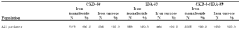
Treating iron deficiency in subjects at risk of cardiovascular adverse events and iron for the management of atrial fibrillation
PatentWO2020144667A1
Innovation
- Administering iron isomaltoside, a high-dose intravenous iron carbohydrate complex, to patients at risk of cardiovascular adverse events, including those with a history of myocardial infarction, stroke, atrial fibrillation, congestive heart failure, and chronic kidney disease, to improve iron stores and reduce cardiovascular risk, combined with other medications to enhance therapeutic benefits.
Regulatory Framework for ECG Devices
The regulatory framework for ECG devices plays a crucial role in ensuring the safety, efficacy, and quality of these medical devices. In the United States, the Food and Drug Administration (FDA) is responsible for overseeing the regulation of ECG devices. These devices are typically classified as Class II medical devices, which require a 510(k) premarket notification submission to demonstrate substantial equivalence to a legally marketed predicate device.
The FDA has established specific guidelines for ECG devices, including performance standards, labeling requirements, and quality system regulations. Manufacturers must comply with these regulations throughout the device lifecycle, from design and development to production and post-market surveillance. The FDA's guidance document "ECG-Related Devices" provides detailed information on the agency's expectations for these devices.
In the European Union, ECG devices fall under the Medical Device Regulation (MDR), which came into effect in May 2021. The MDR introduced more stringent requirements for medical device manufacturers, including enhanced clinical evaluation processes and post-market surveillance. ECG devices are typically classified as Class IIa or IIb devices under the MDR, depending on their specific intended use and risk profile.
The International Electrotechnical Commission (IEC) has developed several standards relevant to ECG devices, such as IEC 60601-2-25 for electrocardiographs and IEC 60601-2-47 for ambulatory electrocardiographic systems. These standards provide detailed technical specifications and safety requirements that manufacturers must adhere to in order to ensure device performance and patient safety.
Regulatory bodies also require manufacturers to implement robust quality management systems, such as those outlined in ISO 13485, to ensure consistent production of safe and effective devices. Additionally, manufacturers must conduct post-market surveillance and report adverse events to regulatory authorities, contributing to the ongoing safety evaluation of ECG devices.
As technology advances, regulatory frameworks continue to evolve to address new challenges and opportunities in ECG device development. For instance, the increasing integration of artificial intelligence and machine learning algorithms in ECG analysis has prompted regulatory bodies to develop guidance on the validation and regulation of these software-based technologies.
P Wave in Personalized Cardiac Care
The P wave, a crucial component of the electrocardiogram (ECG), represents atrial depolarization and plays a significant role in personalized cardiac care. As healthcare continues to evolve towards more individualized approaches, understanding the impact of P waves on atrial electrical activity becomes increasingly important for tailoring treatments and interventions to each patient's unique cardiac profile.
P wave analysis provides valuable insights into atrial conduction patterns and potential abnormalities. By examining P wave morphology, duration, and amplitude, clinicians can identify early signs of atrial pathologies, such as atrial enlargement, conduction delays, or ectopic foci. This information is instrumental in developing personalized treatment strategies and monitoring disease progression.
In the context of personalized cardiac care, P wave analysis can be utilized to assess the risk of atrial fibrillation (AF), a common cardiac arrhythmia. P wave duration and dispersion have been shown to be predictive markers for AF development. By incorporating these parameters into risk assessment models, healthcare providers can identify high-risk individuals and implement preventive measures tailored to their specific cardiac profile.
Furthermore, P wave characteristics can guide the selection and optimization of antiarrhythmic therapies. Different drugs may have varying effects on atrial conduction, and P wave analysis can help clinicians evaluate the efficacy of treatments and adjust dosages accordingly. This personalized approach to medication management can improve outcomes and reduce adverse effects.
In the realm of cardiac device therapy, P wave analysis is crucial for optimizing pacemaker and defibrillator settings. By accurately detecting and interpreting P waves, these devices can provide more physiological pacing and better discriminate between supraventricular and ventricular arrhythmias. This leads to improved device performance and enhanced patient safety.
Advanced signal processing techniques and machine learning algorithms are now being applied to P wave analysis, enabling more sophisticated interpretation of atrial electrical activity. These technologies can detect subtle changes in P wave morphology that may not be apparent to the human eye, potentially uncovering new biomarkers for personalized risk stratification and treatment selection.
As we move towards an era of precision medicine, the integration of P wave analysis with other cardiac biomarkers and genetic information holds promise for even more personalized cardiac care. This holistic approach can lead to more accurate diagnosis, targeted therapies, and improved patient outcomes in the management of atrial arrhythmias and other cardiac conditions.
Unlock deeper insights with Patsnap Eureka Quick Research — get a full tech report to explore trends and direct your research. Try now!
Generate Your Research Report Instantly with AI Agent
Supercharge your innovation with Patsnap Eureka AI Agent Platform!