Method and device for automatic distinguishing contrast agents in bone or calcium containing substance and soft tissue

A technology of contrast agents and substances, applied in image analysis, equipment for radiological diagnosis, image data processing, etc., can solve problems such as bone interference, and achieve the effect of high differentiation reliability

Inactive Publication Date: 2007-10-03
SIEMENS AG
View PDF0 Cites 22 Cited by
  • Summary
  • Abstract
  • Description
  • Claims
  • Application Information

AI Technical Summary

Problems solved by technology

But other body components such as bones can interfere when viewing this CT image because bones also absorb x-rays strongly

Method used


Image

  • Method and device for automatic distinguishing contrast agents in bone or calcium containing substance and soft tissue
  • Method and device for automatic distinguishing contrast agents in bone or calcium containing substance and soft tissue

Examples


Embodiment Construction

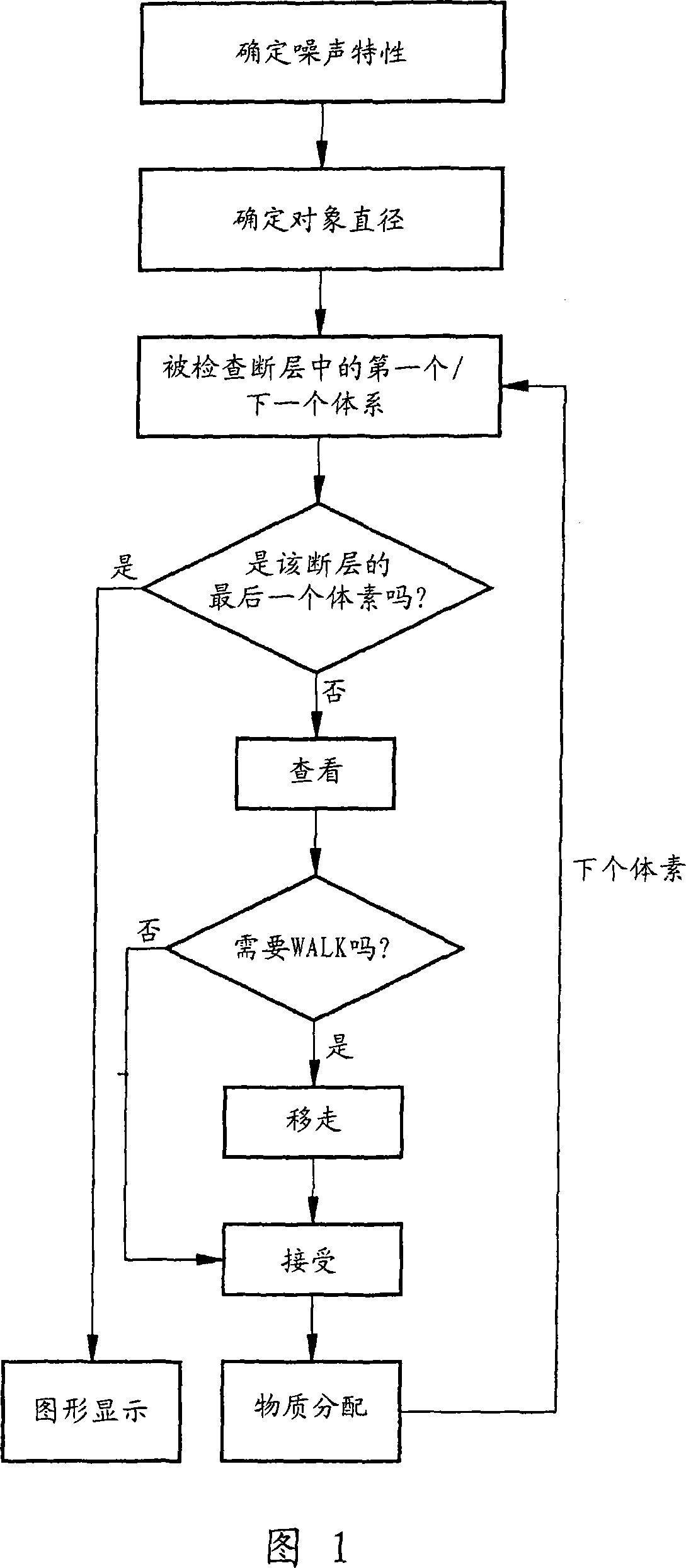
[0028] In the following example, a dual-energy computed tomography device is used to perform a dual-energy CT scan of a subject, in which raw data are obtained at two different X-ray energies at the same time. The different X-ray energies are obtained by the different tube voltages of the X-ray tubes used, in this example 80 kV and 140 kV. The two CT images are independently reproduced from the original data by a well-known reproduction algorithm. Each of the two image data groups obtained here includes the corresponding HU value at each X-ray energy for each voxel of the examination body.

[0029] Here, it should be ensured regardless of the data drawing and the computer tomography equipment used. When the body substance to be distinguished appears or is located at different positions of the subject, the HU value of the body substance is stable to a certain extent. of. But this is a given for most of the computer tomography equipment available on the market.

[0030] The examples...


PUM


Abstract

The invention relates to a method and a device for automatically differentiating bones or other calcium-containing materials and contrast agent in soft tissue of an object area. In at least one embodiment of the method, two image data records of two computed tomography pictures of the object are provided that have been recorded in the context of a different spectral distribution of the X-radiation. For voxels of at least one interesting slice, there is calculated from the two image data records a ratio r that is yielded from measured or averaged X-ray attenuation values of the respective voxel or its environment in the context of the different spectral distributions of the X-radiation and prescribed X-ray attenuation values of soft tissue in the context of the different spectral distributions of the X-radiation according to a prescribed calculation rule. Upon overshooting of a threshold value for the ratio r the respective voxel is assigned either contrast agent or calcium-containing material as a function of the magnitude of r. The proposed method and / or the associated device enable a reliable automatic differentiation of bones or other calcium-containing materials and contrast agent in computed tomography pictures.

Description

Technical field [0001] The present invention relates to a method and device for automatically distinguishing bones or other calcium-containing substances from contrast agents in soft tissues of a target part, wherein two computed tomography of the target part are recorded under different X-ray spectrum distributions Radiography, and reproduce two image data sets of the target part from the raw data of the two computed tomography images. These image data sets contain the X-ray attenuation of the voxel of the target part under each X-ray spectrum distribution value. Background technique [0002] In many examinations with the aid of computed tomography, a contrast agent is injected into the patient, so that the blood vessels can be particularly clearly identified in the obtained CT images. However, other body components such as bones may interfere when observing the CT image, because bones also strongly absorb X-rays. Therefore, this area is sometimes removed from the image by calcu...

Claims


Application Information

Patent Timeline
Patent Type & Authority Applications(China)
IPC IPC(8): A61B6/03G06T7/00G06F19/00
CPCG06T7/0012A61B6/4014A61B6/4035A61B6/482A61B6/505G06T7/0081G06T2207/10081G06T2207/20216G06T2207/30008G06T7/11
Inventor 伯恩哈德·克劳斯迈克尔·格拉斯拉克
Owner SIEMENS AG
Why Eureka
Try Eureka