A method and device for calculating the proportion of inflammation and necrosis in medical images

A calculation method and technology of medical images, applied in the fields of medicine and image recognition, to reduce the difficulty of convergence

Active Publication Date: 2022-07-22
BEIJING FRIENDSHIP HOSPITAL CAPITAL MEDICAL UNIV
View PDF0 Cites 0 Cited by
  • Summary
  • Abstract
  • Description
  • Claims
  • Application Information

AI Technical Summary

Problems solved by technology

[0003] The purpose of the embodiment of the present invention is to provide a method and device for calculating the proportion of inflammation and necrosis in medical images. problems, and reduce the difficulty of convergence, quickly and accurately obtain the proportion of inflammation and necrosis

Method used


Image

  • A method and device for calculating the proportion of inflammation and necrosis in medical images
  • A method and device for calculating the proportion of inflammation and necrosis in medical images
  • A method and device for calculating the proportion of inflammation and necrosis in medical images

Examples


Embodiment Construction

[0031] The specific implementations of the embodiments of the present invention will be described in detail below with reference to the accompanying drawings. It should be understood that the specific implementation manners described herein are only used to illustrate and explain the embodiments of the present invention, and are not used to limit the embodiments of the present invention.

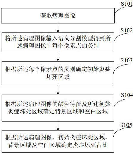
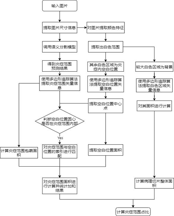
[0032] figure 1 It is a schematic flowchart of a method for calculating the proportion of inflammation and necrosis in medical images of the present invention, such as figure 1 As shown, step S101 is to acquire a pathological image, the pathological image includes inflammation and necrosis foci, and the pathological image is a pathological slice image.

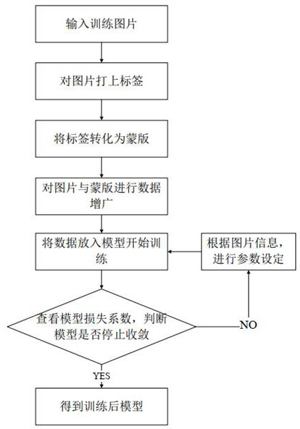
[0033] Step S102 is to input the pathological image into a semantic segmentation model to obtain the category of each pixel in the pathological image, including: acquiring a feature set of the pathological image; performing feature fusion o...


PUM


Abstract

Embodiments of the present invention provide a method and device for calculating the proportion of inflammation and necrosis in a medical image. The calculation method includes acquiring a pathological image; inputting the pathological image into a semantic segmentation model to obtain the category of each pixel in the pathological image ; Determine the initial inflammatory necrosis area according to the category of each pixel point; Determine the background area and blank area according to the color feature of the pathological image and the initial inflammatory necrosis area; According to the pathological image, the initial inflammatory necrosis area, the background The area and blank area were used to determine the proportion of inflammation and necrosis. The calculation method solves the problem of nesting between inflammation and necrosis, reduces the difficulty of convergence, and obtains the proportion of inflammation and necrosis quickly and accurately.

Description

technical field [0001] The invention relates to the fields of medicine and image recognition, in particular to a method and device for calculating the proportion of inflammation and necrosis in medical images. Background technique [0002] In existing medical practice, medical personnel mainly judge the severity of a patient's disease by analyzing the coverage of inflammation and necrosis in pathological sections. Therefore, in medical clinical diagnosis, the rapid and accurate judgment of the proportion of inflammation and necrosis is particularly important. The existing method mainly uses the traditional manual method to manually delineate the site of inflammation and necrosis and calculate the delineated range. This method is more efficient for cases with a small number of inflammation and necrosis, but it is not suitable for large-scale and complex pathological slice images. Said that due to the variable size of inflammatory necrosis and the nesting of inflammatory necr...

Claims


Application Information

Patent Timeline
Patent Type & Authority Patents(China)
IPC IPC(8): G06V10/26G06K9/62G06T7/11G06T7/62G06T7/90G06V10/764G06V10/80G06V20/70
CPCG06T7/11G06T7/62G06T7/90G06F18/24G06F18/253
Inventor 林旭辉王昱赵新颜
Owner BEIJING FRIENDSHIP HOSPITAL CAPITAL MEDICAL UNIV
Why Eureka
Try Eureka