Endoscope apparatus

a portable, endoscope technology, applied in the field of medical devices, can solve the problems of inconvenient carrying of the endoscope, the surface of the internal organs is delicate and susceptible to damage, and the conventional endoscope is an expensive apparatus, so as to prevent cross-contamination between patients subject to the endoscope examination, the portable endoscope apparatus is convenient to carry, and the effect of convenient portability

Inactive Publication Date: 2007-02-15
TIEN DER YANG
View PDF11 Cites 17 Cited by
  • Summary
  • Abstract
  • Description
  • Claims
  • Application Information

AI Technical Summary

Benefits of technology

[0007] In order to solve the above drawbacks in the prior art, a primary objective of the present invention is to provide an endoscope apparatus, allowing the endoscope to be wound and received in a body of the apparatus, wherein the body is provided with a carrying member to allow a user to easily carry the endoscope apparatus that is thus more convenient to use.
[0008] Another objective of the invention is to provide an endoscope apparatus, which is formed with a head portion for capturing images, and a rotatable lens with a adjustable focal length in the head portion, to allow images to be accessed and clearly taken.
[0009] Still another objective of the invention is to provide an endoscope apparatus, which is formed with a biologically compatible and disposable cover for encapsulating a surface of the endoscope in contact with body cavities or canals of a patient, and a clamping member for holding the cover in position so as to avoid displacement of the cover when in contact with walls of the body cavities or canals and thereby protect the patient against cross-contamination.
[0011] In order to achieve the foregoing and other objectives, the present invention provides an endoscope apparatus, comprising: (1) a head portion comprising (a) an image capturing unit for capturing images, and (b) an illuminator module for providing light to the image capturing unit, wherein the image capturing unit comprises (i) a rotatable lens with an adjustable focal length, and (ii) a converting module for converting the images captured by the image capturing unit into digital image data; (2) a flexible connecting member connected to the head portion to form an endoscope, wherein the connecting member transmits power to the head portion for performing the image capturing, and also transmits the captured images from the head portion; and (3) a body for receiving the head portion and the connecting member therein, the body comprising (a) a carrying member for allowing a user to carry the endoscope apparatus via the carrying member, (b) a display unit for displaying the images captured by the image capturing unit, (c) a socket for accommodating a storage medium for storing data, (d) a power supply module for providing power to the endoscope apparatus, (e) an out port for being connected to an external data processor and performing data transmission, and (f) a control interface for adjusting an operating mode of the image capturing unit. The endoscope apparatus provided in the invention comprises an endoscope consisting of the head portion and the flexible connecting member to examine the condition of an internal body cavity of a human. The connecting member comprises a plurality of optical fibers and a cladding for covering the optical fibers, and is used as a transmission pathway for transmitting image data and control signals. The lens provided in the head portion can be adjusted to a proper focal length and rotated in position according to the user setting so as to obtain sharp images of the body cavity and even at blind angle positions. Moreover, since the optical fibers are flexible, the connecting member can be rolled up and received in the body. Furthermore, the carrying member provided on the body allows the user to conveniently carry the endoscope apparatus, making the endoscope apparatus portable and easy to use.
[0012] In addition, the endoscope apparatus further comprises a disposable cover and a clamping member, wherein the cover is used to cover part of the endoscope (including the head portion and part of the connecting member) that is to be inserted into the body cavity, and the clamping member is to fix the cover in place on the connecting member, such that cross-contamination between patients subject to the endoscope examination can be prevented. The images of the body cavity captured by the endoscope apparatus can be converted into image files and then transmitted to the external data processor where the image data are analyzed and integrated. Subsequently, the processed image files can be transmitted to a distant terminal such as medical center from the external data processor via a network connected to the data processor so as to perform a further analysis of the image files to give more thorough and accurate diagnosis.

Problems solved by technology

Insertion of endoscope into the body cavity may often discomfort a patient, even leading to shock in a severe case.
Further, surfaces of the internal organs are delicate and might be wounded by the endoscope.
Moreover, the conventional endoscope is an expensive apparatus, which needs to be cleaned and sterilized before the next use.
However, since the endoscope is complex in structure, and the cleaning equipment for the endoscope is expensive, in case the standard cleaning procedure is not perfectly followed by an operator who is not familiar or careful with the cleaning equipment, the endoscope fails to be thoroughly and properly cleaned and sterilized, making patients who have ever been examined using this contaminated endoscope be in danger of cross-contamination.
In addition, remote areas such as rural areas are often short of medical supplies and can only rely on crude medical devices for medical examination and disease diagnosis, such that patients at these remote rural areas are usually not subject to good and timely treatments, making the patients' condition even worse.
Although the government and private organizations periodically organize medical teams to provide more advanced medical devices and medicines to the remote areas, the effects achieved thereby on improvement in the medical condition of the remote areas are limited.
A patient from the remote areas may require a detailed examination on his internal body at a large hospital having advanced and expensive examining devices such as an endoscope.
However, these same devices are usually large in size and cannot be carried with the medical teams to the remote areas.
Another important issue is the lack of medical human resources in the remote areas to give a correct diagnosis from image data taken by the endoscope even if it is brought to the remote areas.

Method used


Image

  • Endoscope apparatus
  • Endoscope apparatus
  • Endoscope apparatus

Examples


Embodiment Construction

[0022] The preferred embodiment of an endoscope apparatus proposed in the present invention is described in detail with reference to FIGS. 1-5.

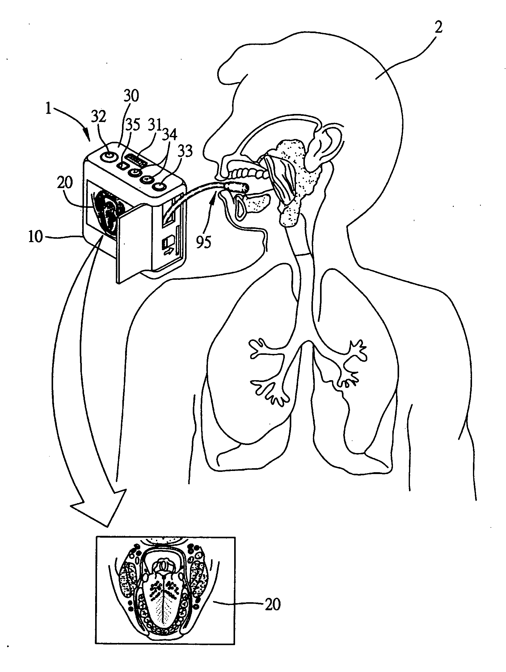
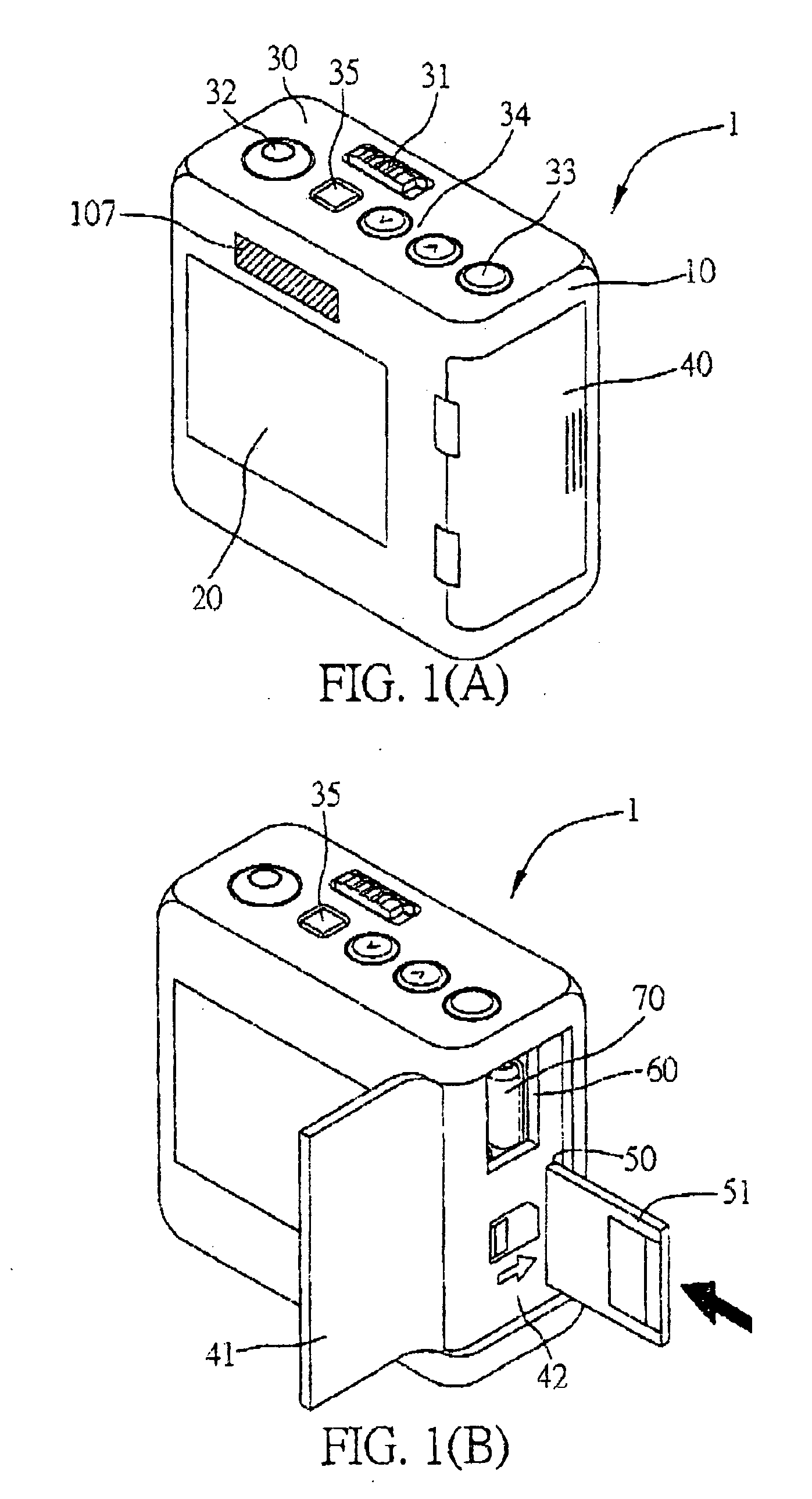
[0023] FIGS. 1(A) and 1(B) show the appearance of the endoscope apparatus 1 according to the present invention. As shown in FIG. 1(A), a body 10 of the endoscope apparatus 1 is provided with a display unit 20 such as liquid crystal display (LCD) screen for displaying images; a control interface 30 for providing operational functions; and a receiving unit 40 with a liftable cap, for receiving part of functioning components of the endoscope apparatus 1. On the control interface 30, there are provided a focal length controller 31 for adjusting a focal length of a lens (not shown) installed in a head portion 70 of the endoscope apparatus 1; an angle controller 32 for adjusting an angle of observation for the lens; a function switch button 33; a selector 34 and a retract button 35. The function switch button 33 allows the function items listed on...


PUM


Abstract

An endoscope apparatus includes a head portion for capturing images, a flexible connecting member made of optical fibers, and a body. The connecting member is used to transmit image data and control signals. A lens is provided in the head portion and adjustable to obtain sharp and clear images. The images are converted into image files and transmitted to an external data processor for data analysis. The optical fibers are flexible, such that the connecting member can be rolled and received in the body when it is not in use. A carrying member is provided on the body to allow a user to conveniently carry the endoscope apparatus. A disposable cover is used to cover the head portion and part of the connecting member that are to be in contact with an internal body cavity of a human, and a clamping member is provided to fix the cover in place.

Description

[0001] This application is a continuation-in-part of application Ser. No. 10 / 886,153 filed Jun. 14, 2004.FIELD OF THE INVENTION [0002] The present invention relates to medical devices for examining internal body cavities, and more particularly, to a portable endoscope apparatus with an adjustable focal length, which is capable of capturing and transmitting image data through optical fibers. BACKGROUND OF THE INVENTION [0003] A conventional endoscope is made of a specialized and elongated tube, consisting of an imaging capturing device and a power supply. With the endoscope being connected to a screen, it allows internal body structures to be displayed on the screen and examined by the doctor for disease diagnosis when the endoscope is inserted into a body cavity. The organs having communication with the outside the body can be examined using the endoscope. For example, the endoscope is inserted through the mouth to the alimentary canal to examine the esophagus, stomach, and duodenum...

Claims


Application Information

Patent Timeline
Patent Type & Authority Applications(United States)
IPC IPC(8): A61B1/00A61B1/04A61B1/227A61B1/233A61B1/24A61B1/303A61B1/307A61B1/31G02B23/24H04N5/225H04N7/18
CPCA61B1/00105A61B1/042A61B1/00052G02B23/2484H04N7/183A61B1/24H04N23/555
Inventor TIEN, DER-YANG
Owner TIEN DER YANG
Why Eureka
Try Eureka