
Fluorescence labeled human triple-negative breast cancer osseous metastasis cell line
A triple-negative breast cancer, fluorescent labeling technology, applied in the field of bioengineering, can solve the problems of the inability to visualize and trace the bone metastasis cell line, and achieve the effect of bone metastasis ability and bone metastasis specificity.
Patent Information
- Authority / Receiving Office
- CN · China
- Patent Type
- Applications(China)
- Current Assignee / Owner
- Publication Date
- 2020-08-28
Smart Images
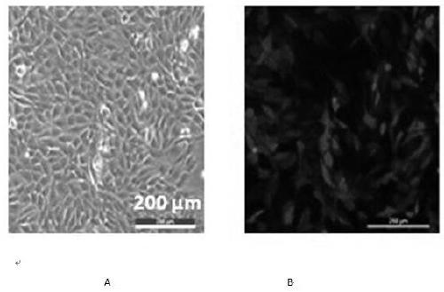
Figure 1 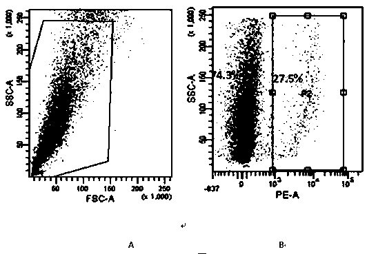
Figure 2 
Figure 3
Abstract
Description
technical field
[0001] The invention belongs to the field of bioengineering and relates to a cell line, in particular to a fluorescence-labeled human triple-negative breast cancer bone metastasis cell line. Background technique
[0002] Since the beginning of this century, the global incidence of breast cancer has increased rapidly, becoming the number one killer of urban women. Although the early diagnosis of breast cancer and the improvement of surgical techniques and radiotherapy and chemotherapy have reduced the mortality rate of breast cancer, the mortality rate of malignant breast cancer accompanied by lung, bone, brain and other organ metastasis is still high. According to statistics, about 70% of uncured breast cancers will have lung metastases, about 60% will have bone and liver metastases, and about 15% will have brain metastases.
[0003] In particular, triple-negative breast cancer accounts for about 15% of breast cancers. It is highly malignant and prone to inv...
Examples
Embodiment 1
[0028] The present invention provides a fluorescently labeled human triple-negative breast cancer bone metastases cell line, which is prepared by the following method:
[0029] (1) Culture parental human triple negative breast cancer cell MDA-MB-231 (purchased from ATCC);
[0030] (2) Amplify the lentiviral vector pLemiR (product of Open biosystems) containing the red fluorescent protein RFP. Using the method of transfecting cells with lentivirus, transfect MDA-MB-231 cells, and prepare MDA-MB-231 cells containing red fluorescent markers;
[0031] (3) Sorting by flow cytometry to obtain red fluorescent RFP-MDA-MB-231 cells ( figure 1 A: white light image of RFP-MDA-MB-231 cells, figure 1 B: fluorescence image of RFP-MDA-MB-231 cells);
[0032] (4) Transplant 1 million RFP-MDA-MB-231 cells into the fourth pair of mammary fat pads in nude mice. Tumors began to form after one week, and the tumor diameter reached 0.8-1.2cm after four to six weeks;
[0033] (5) Nude mice were k...
Embodiment 2
[0041] The MDA-MB-231-BM cells obtained in the present invention are tested for cell proliferation, cell migration, bone metastasis and tumorigenesis, and the gene expression characteristics of the MDA-MB-231-BM cells are tested. The experimental steps are as follows:
[0042] 1: Colony formation assay was used to detect cell proliferation ability.
[0043] 1) After digestion of MDA-MB-231 and MDA-MB-231-BM in good condition in logarithmic growth phase, add complete medium to make cell suspension;
[0044] 2) Count the cells of the cell suspension, inoculate 2,000 cells in a 12-well plate, add 1 mL of complete medium to the 12-well plate, and spread the cells evenly on the culture plate;
[0045] 3) Place the 12-well plate in the cell incubator, and change the medium every 2-3 days after the cells adhere to the wall to avoid contamination;
[0046] 4) When a round cell mass can be seen under the microscope (about the 10th day), take out the 12-well plate, discard the culture...