
Plaque Removal and Differentiation of Tooth and Gum
a technology of tooth and gum, which is applied in the field of tooth and gum plaque removal and differentiation, can solve the problems of deep pockets between teeth and gums, loss of bone that holds teeth in place, and noisy pumps in these units, and contribute to patient discomfor
Patent Information
- Authority / Receiving Office
- US · United States
- Patent Type
- Applications(United States)
- Current Assignee / Owner
- Publication Date
- 2011-12-22
Smart Images
Figure 1 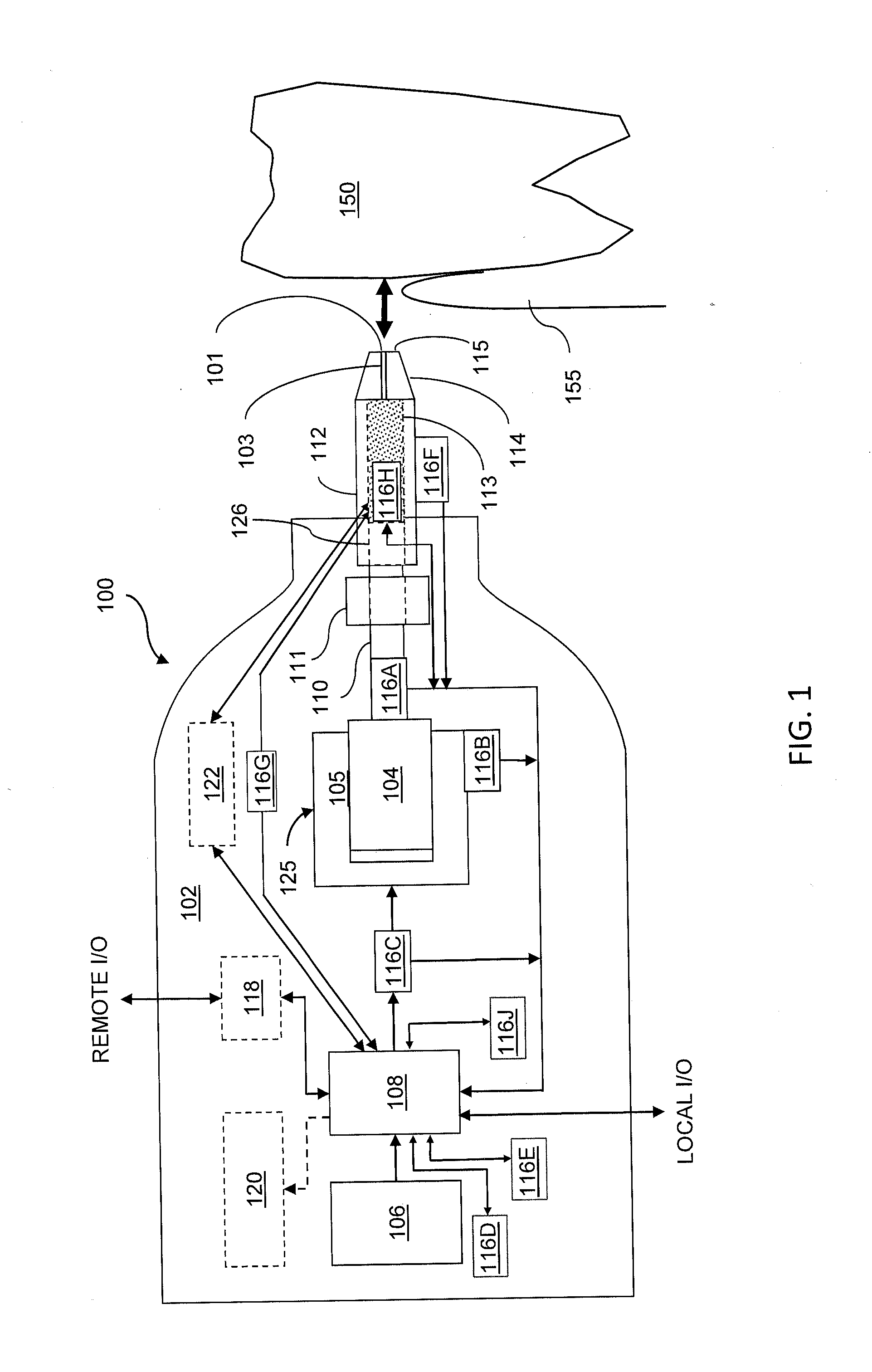
Figure 2 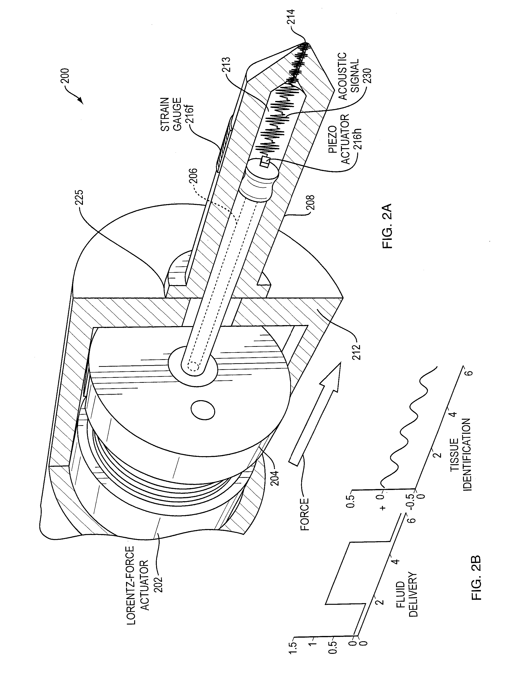
Figure 3
Abstract
Description
RELATED APPLICATIONS
[0001] This application claims the benefit of U.S. Provisional Application No. 61 / 286,632, filed on Dec. 15, 2009, and U.S. Provisional Application No. 61 / 286,651, filed on Dec. 15, 2009. The entire teachings of the above applications are incorporated herein by reference.BACKGROUND OF THE INVENTION
[0002] In the oral cavity, indigenous bacteria are often associated with two major oral diseases; caries and periodontitis [H. Marcotte and M. C. Lavoie, Oral microbial ecology and the role of salivary immunoglobulin A, Micro Mol Bio 62 (1998) 71-109]. The diverse structures within the mouth (i.e. the tooth surface, subgingival space, and tongue) support several different microbial communities. The supragingival environment of the oral cavity is regulated by saliva, a complex mixture of water, electrolytes (e.g. sodium, potassium, calcium, chloride, magnesium, bicarbonate, phosphate), enzymes (e.g. lysozyme, lactoferrin, peroxidase), proteins (e.g. sIgA, glycoproteins etc...
Examples
Embodiment Construction
"d_n">[0024]FIGS. 5A-5D are perspective diagrams showing delivery of water and black beads to the gum line and sulcus;
[0025]FIG. 6 is a top perspective diagram of an exemplary detector for detecting an analyte from a sample collected in a disposable tube;
[0026]FIGS. 7A and 7B are schematic block diagrams of a needle-free transport device providing sampling and analysis capability, respectively shown in the sampling and fluid ejection configurations;
[0027]FIG. 8 is a top perspective diagram of an exemplary detector for detecting an analyte using a polymer strip;
[0028]FIG. 9A is a schematic diagram showing binding of an analyte (antibody) to a solid support (polypyrrole film strip);
[0029]FIG. 9B is a graph of light absorbance versus antigen concentration;
[0030]FIG. 10 is a schematic diagram showing a detector for detecting a marker or analyte of oral health status.
DETAILED DESCRIPTION OF THE INVENTION
[0031]A description of example embodiments of the invention follows.
[0032]The entire ...