
Breast cancer prognosis
Patent Information
- Authority / Receiving Office
- US · United States
- Current Assignee / Owner
- Publication Date
- 2013-11-28
Smart Images
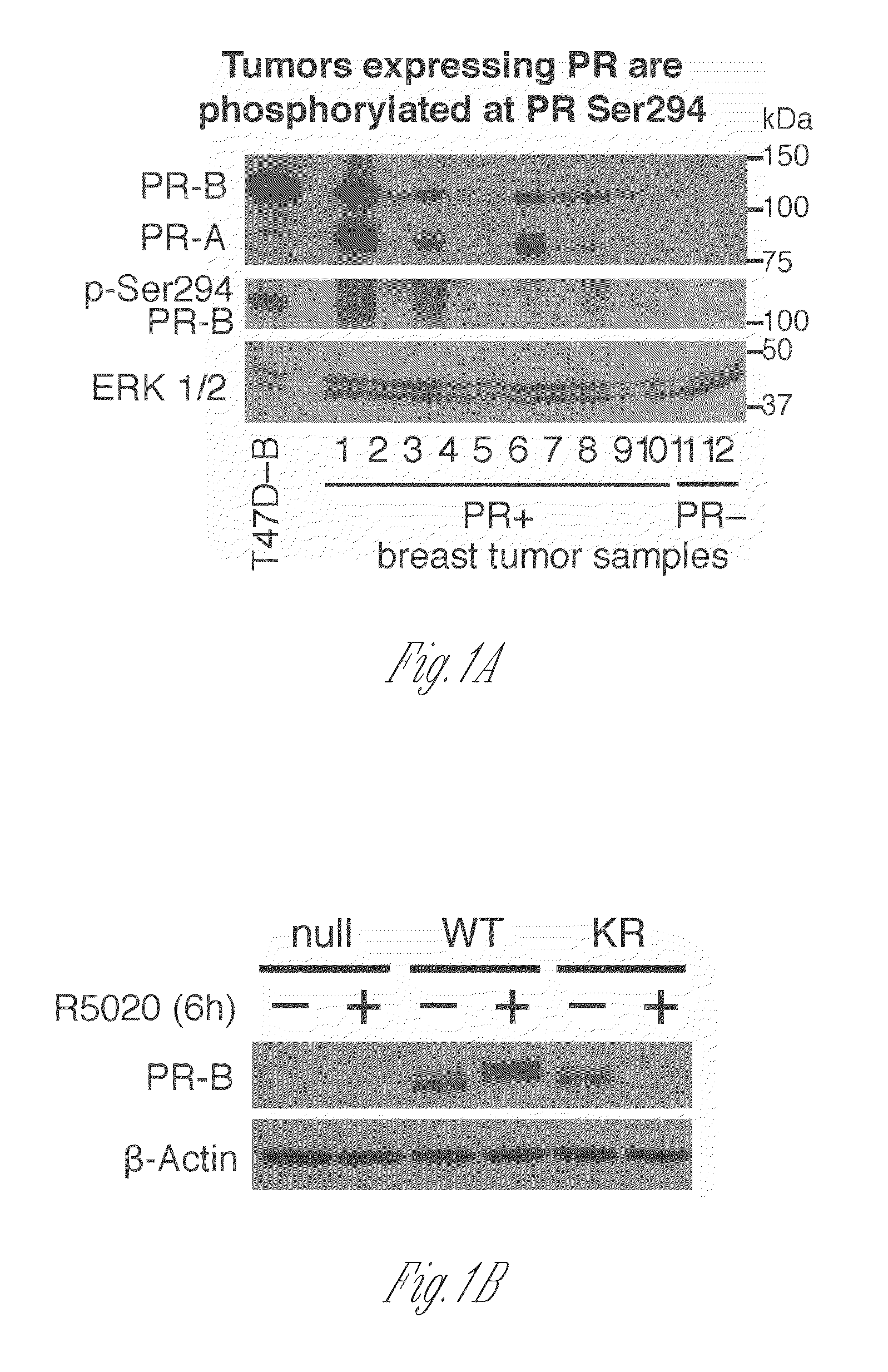
Figure 1 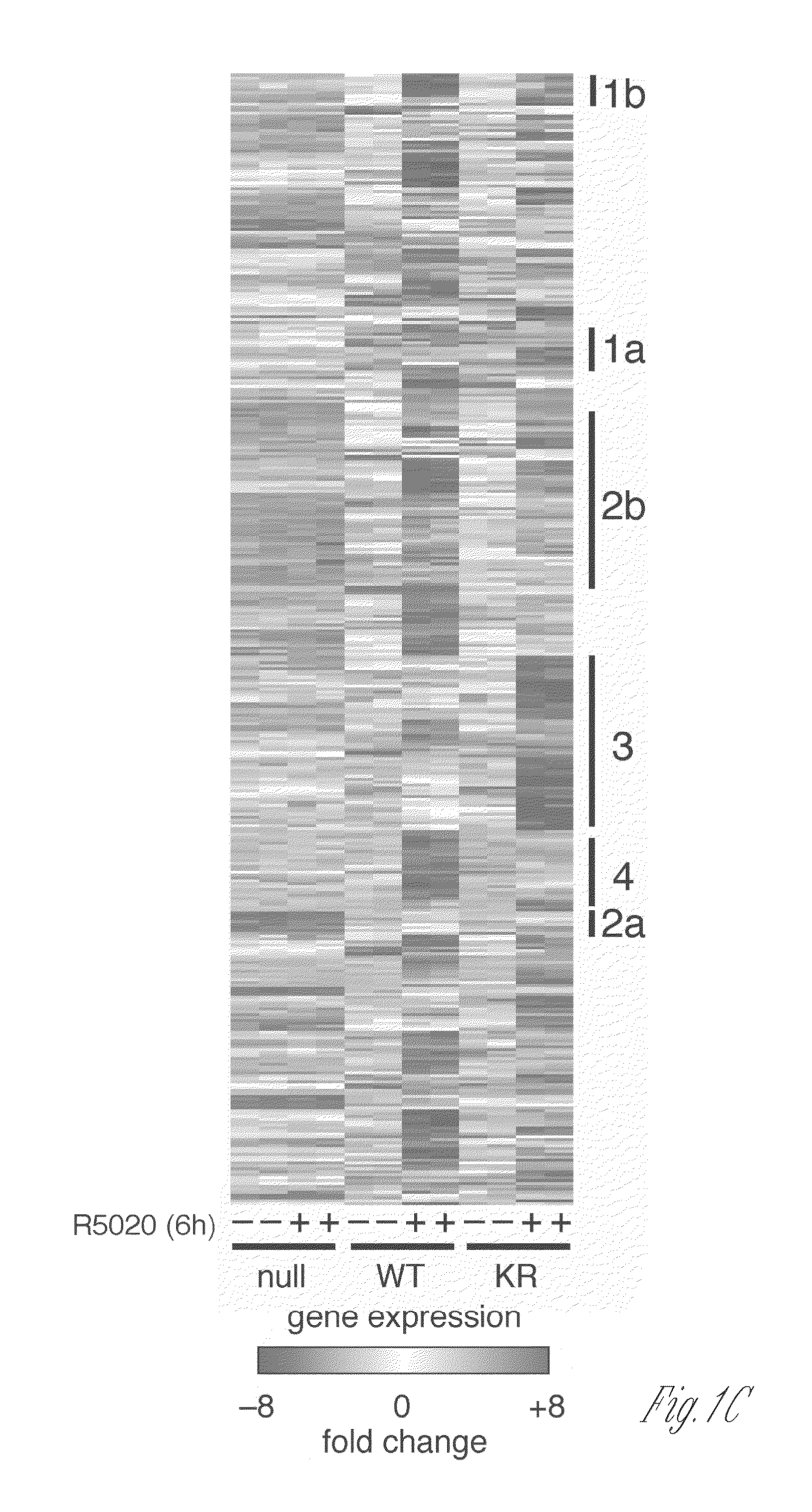
Figure 2 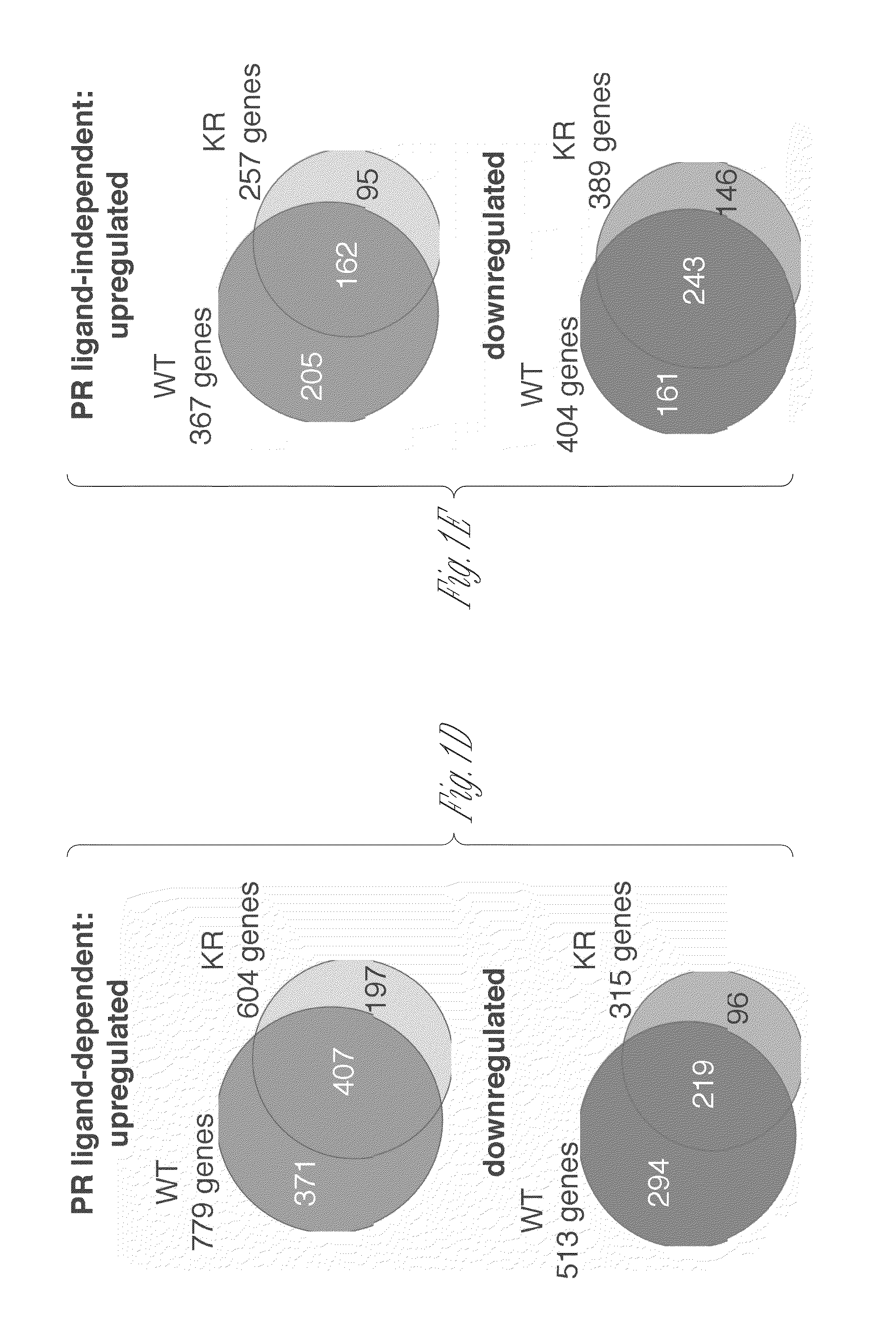
Figure 3
Abstract
Description
CROSS-REFERENCE TO RELATED APPLICATIONS
[0001] This application claims priority from U.S. Provisional Application No. 61 / 639,407, filed Apr. 27, 2012, the entire disclosure of which is herein incorporated by reference.STATEMENT OF GOVERNMENT RIGHTS
[0002] This invention was made with the assistance of government support under United States Grant No. CA1159712-01 from the National Institutes of Health. The government has certain rights in the invention.BACKGROUND OF THE INVENTION
[0003] Current mRNA (e.g., gene expression based) prognostic breast cancer screening tests (such as Oncotype DX) assay for expression of a limited number of unrelated genes, each known to be involved in breast cancer progression. Because breast cancer is a very heterogeneous disease, these tests fail to select those patients who are most likely to benefit from a given targeted therapy, including a rapidly growing list of existing and new drugs. Thus, health care providers are forced to try random combinations of a...
Examples
examples
[0145]The following examples are provided in order to demonstrate and further illustrate certain embodiments and aspects of the present invention and are not to be construed as limiting the scope thereof.
example i
Phosphorylated and SUMO-Deficient Progesterone Receptors Drive Proliferative Gene Signatures During Breast Cancer Progression
Materials and Methods
Progesterone Receptor Expression in Human Breast Tumor Samples
[0146]De-identified human breast tumor samples were obtained from the University of Minnesota Tissue Procurement Facility's Biological Materials Procurement Network (BioNet) for protein and mRNA analysis. Frozen tissue samples were derived from patients diagnosed with either ductal carcinoma, infiltrating ductal carcinoma, lobular carcinoma, or metastatic carcinoma. Specimens were analyzed by the University of Minnesota clinical pathology department and scored for estrogen receptor (ER) and progesterone receptor (PR) expression using standard clinical histological methods. Tumor samples were harvested individually for protein or mRNA using standard methods (frozen tissue grinding, RIPA buffer, tri-reagent), and total PR, phospho-Ser294 PR, and ERK1 / 2 protein expression levels we...