Check patentability & draft patents in minutes with Patsnap Eureka AI!

Composition for imaging atherosclerosis and method for diagnosing atherosclerosis by using same

A technology of atherosclerosis and composition, which is applied in the direction of drug combination, preparation for in vivo test, nuclear magnetic resonance/magnetic resonance imaging contrast agent, etc., can solve the problem of limited physical conditions, high production cost, atherosclerosis Reduced diagnostic accuracy and other issues, to achieve effective diagnosis, low production cost, and high accuracy

Active Publication Date: 2016-10-26
KOREA UNIV RES & BUSINESS FOUND +1
View PDF6 Cites 1 Cited by
  • Summary
  • Abstract
  • Description
  • Claims
  • Application Information

AI Technical Summary

Problems solved by technology

[0004] However, diagnostic methods for imaging atherosclerosis using F18-FDG have several problems
First, because FDG is a glucose analogue, control of physical conditions for diagnosis is limited due to potential effects on blood glucose or metabolism-related hormones
Therefore, the accuracy of the diagnosis of atherosclerosis is reduced
Furthermore, because fasting or glycemic control is necessary, this approach is limited for high-risk groups such as those with diabetes
Second, although atherosclerosis occurs frequently in the brain and heart, it is difficult to detect atherosclerosis using F18-FDG because they use blood glucose as energy
Again, the production cost is too high, because expensive equipment such as cyclotron is required to prepare F18-FDG

Method used


Image

  • Composition for imaging atherosclerosis and method for diagnosing atherosclerosis by using same
  • Composition for imaging atherosclerosis and method for diagnosing atherosclerosis by using same
  • Composition for imaging atherosclerosis and method for diagnosing atherosclerosis by using same

Examples


Embodiment 1

[0035] Embodiment 1: for Ga-68 ( 68 Preparation of Ga) labeled NOTA-MSA

[0036]

[0037] After dissolving 20 mg of human serum albumin in 5 mL of 0.1 M carbonate buffer (pH 9.5) and adding 5.5 mg of α-L-isothiocyanatophenyl mannopyranoside, the The reaction was carried out with stirring for 20 hours. Then, the reaction solution was stored at -70°C.

[0038]

[0039] After adding 10 mg of p-SCN-Bz-NOTA to 1 mL of mannosylated human serum albumin (13.6 mg / mL) prepared in step 1, the reaction was carried out at room temperature for 1 hour. After the reaction, benzyl NOTA and phenylmannose-conjugated human serum albumin were separated and purified with a Sephadex G-25 column.

Embodiment 2

[0040] Example 2: Preparation of kits for imaging mannose receptors

[0041] Add 1 mL of benzyl NOTA and phenylmannose-conjugated human serum albumin (13.6 mg / mL) to 0.3 mL of sodium acetate buffer (0.5 M, pH 5.5) and transfer an amount equivalent to 1 mg of protein After filling each vial, the mixture was lyophilized and stored at -70°C.

Embodiment 3

[0042] Example 3: Preparation using a kit for imaging mannose receptors 68 Ga-labeled compounds

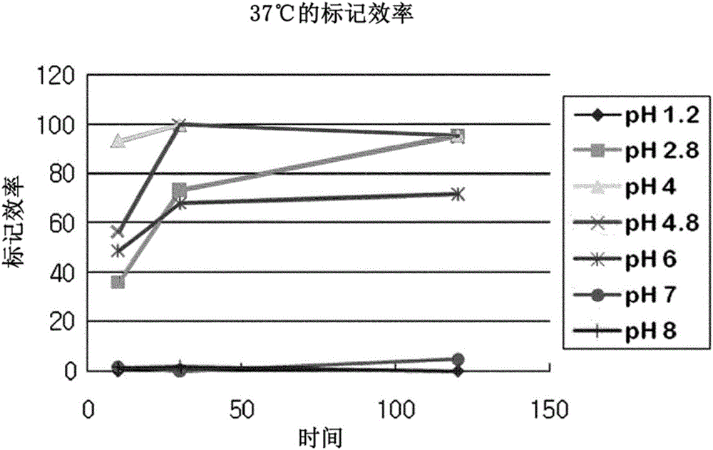
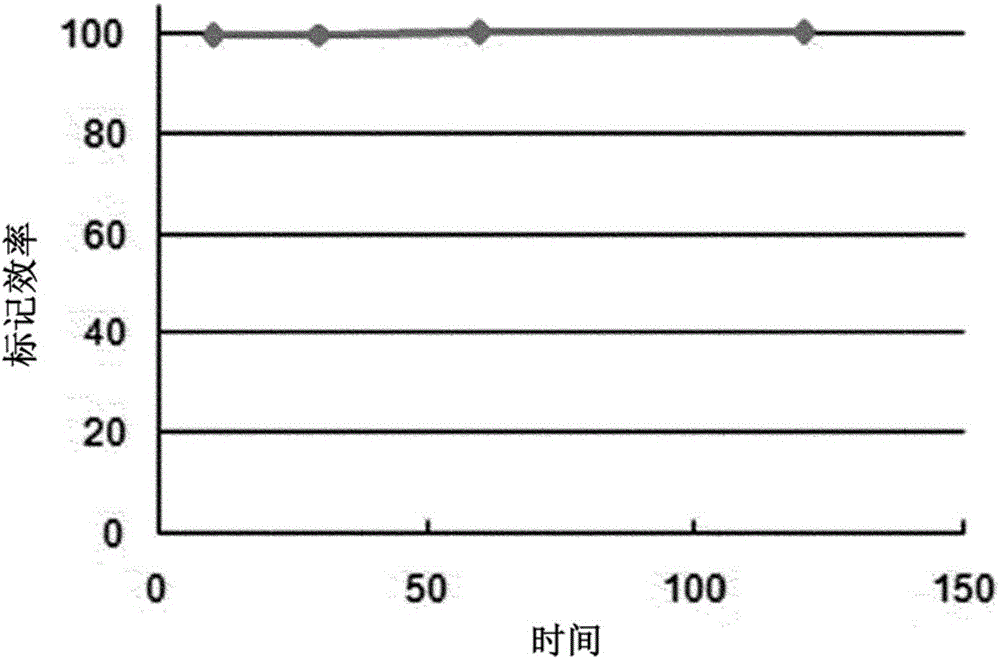
[0043] When adding to the kit of embodiment 2 use 68 Ge / 68 1 mL prepared by a Ga generator (Cyclotron Co., Russia) 68 After GaCl in 0.1 M hydrochloric acid solution, the labeling efficiency was measured later by TLC for 10 minutes, 30 minutes, 1 hour and 2 hours while the reaction was carried out at 37°C. ITLC-SG (Gelman Co., USA) was used as a stationary phase, and 0.1M citric acid solution was used as a mobile phase. The distribution of radioactivity on the ITLC plate was measured using a TLC scanner (Bioscan Co.). marked 68 Ga remains at the origin and unlabeled 68 Ga moves to the solvent front ( figure 1 ). Labeling was almost complete after 30 minutes at 37°C, pH 4-5 ( figure 2 ). After mixing with human serum and incubating at 37°C, the labeled 68 Stability of Ga-NOTA-MSA. The results are shown in image 3 middle. From image 3 It can be seen that the ...


PUM

PropertyMeasurementUnit
sizeaaaaaaaaaa
sizeaaaaaaaaaa

Abstract

The present invention relates to a composition for imaging atherosclerosis and a method for diagnosing atherosclerosis by using the same. The composition for imaging atherosclerosis according to the present invention shows excellent atherosclerosis diagnosis accuracy, enables atherosclerosis diagnosis even for a person with diseases requiring glycemic control such as diabetes, and enables effective diagnosis even for atherosclerosis occurring in the brain and heart. In addition, the manufacturing cost thereof is low compared with a conventional imaging composition for atherosclerosis diagnosis. Therefore, atherosclerosis can be effectively diagnosed by using the same.

Description

technical field [0001] The present disclosure relates to compositions for imaging atherosclerosis and methods of diagnosing atherosclerosis using the compositions. Background technique [0002] Atherosclerosis refers to a disease in which cholesterol deposits on the inside (intima) of blood vessels and intimal cells proliferate at the deposits, resulting in narrowing or blockage of blood vessels and reduced blood flow to the periphery. More specifically, atherosclerosis is a vascular disease in which, just as the diameter of an aging water pipe narrows due to the deposition of rust and impurities, cholesterol penetrates into the intimal layer of the blood vessel, where intimal cells and macrophages proliferate, resulting in Atheroma formation. The nucleus of an atheroma is composed of an acellular lipid core covered by a hard fibrous plaque. When the plaque becomes unstable, it ruptures and forms a blood clot in the blood vessel. Hemorrhage into the atheroma results in ra...

Claims


Application Information

Patent Timeline
Patent Type & Authority Applications(China)
IPC IPC(8): A61K51/04A61K49/06A61K51/02A61P9/10
CPCA61K51/04A61K51/048A61K51/0482A61K51/0491A61P9/10A61K51/088A61K51/081A61K51/08A61K51/044
Inventor 徐汞锡金成恩金应周郑载民
Owner KOREA UNIV RES & BUSINESS FOUND
Features
  • R&D
  • Intellectual Property
  • Life Sciences
  • Materials
  • Tech Scout
  • Unparalleled Data Quality
  • Higher Quality Content
  • 60% Fewer Hallucinations
Learn More