
A method for compound automatic radiotherapy planning
An algorithm and image technology, applied in the field of compound automatic radiotherapy planning, can solve problems such as long calculation time, many manpower and material resources, and inability to be widely used, and achieve the effect of low efficiency
Patent Information
- Authority / Receiving Office
- CN · China
- Patent Type
- Patents(China)
- Current Assignee / Owner
- Publication Date
- 2021-08-27
Smart Images
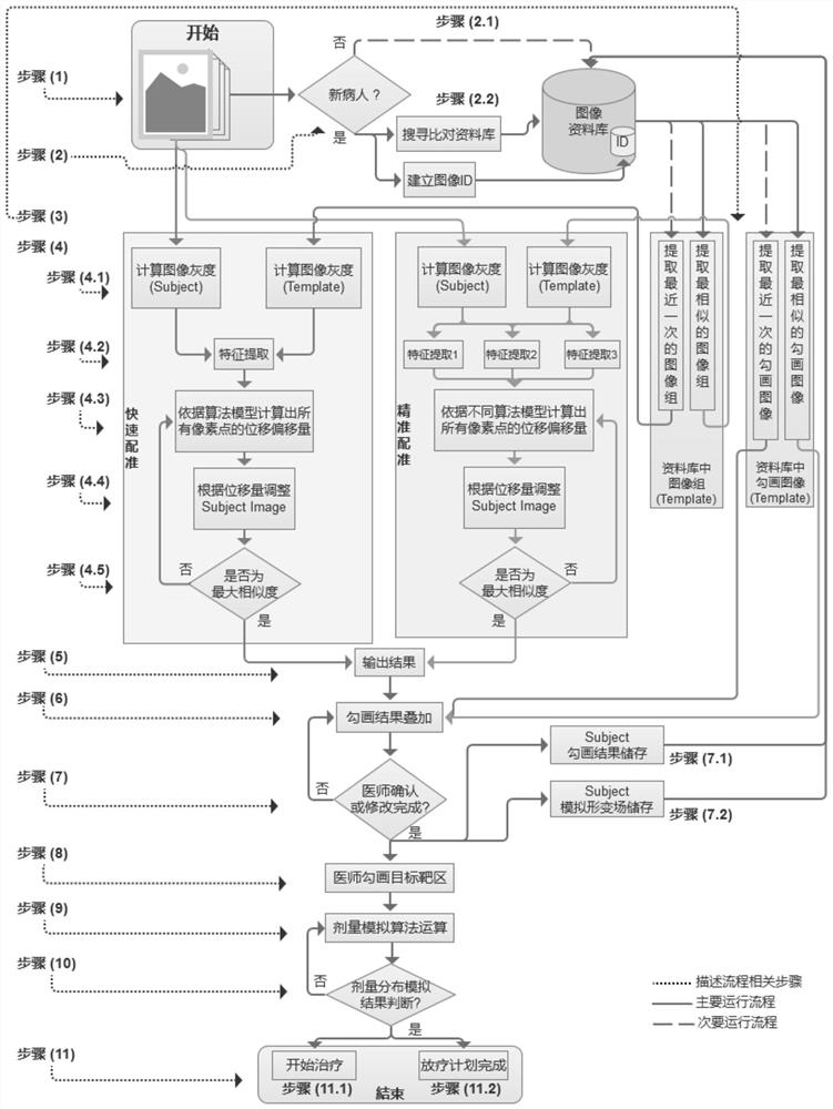
Figure 1 
Figure 2 
Figure 3
Abstract
Description
technical field
[0001] The invention relates to a method for compound automatic radiotherapy planning, in particular to an automatic online compound optimization application method of radiotherapy plan. Background technique
[0002] In the process of radiation therapy (Radiation Therapy), the patient's positioning CT is first scanned, and then the target area is delineated by the doctor and the outline of the organs at risk, and then designed by the physicist or dosimetrist through the commercial Treatment Planning System (TPS) The treatment plan is optimized, and the quality assurance of the treatment plan is carried out by a physicist. Finally, the treatment plan can be used in the subsequent fractionated (Fraction) treatment sessions.
[0003] However, such a treatment modality does not take into account the patient's anatomical changes during subsequent treatment sessions, such as changes in tumor volume and location, changes in patient body contours, changes in gastroin...
Examples
Embodiment 1
[0119] When an untreated tumor patient (Subject) comes to the radiation therapy department, a set of tumor localization CT images will be called out or taken for the patient first, and the doctor will read the relevant medical history and relevant examination reports. The next steps are as follows:
[0120] Step (1): Read patient data, which includes tumor images and image information of surrounding organs and tissues, including CT (Computed Tomography), cone-beam CT (Cone-Beam CT), ultrasound (Ultrasound, US), PET (PositronEmission Tomography) or Magnetic Resonance (Magnetic Resonance, MR) and other images; and information including the patient's basic data and relevant medical history;
[0121] Step (2): Based on the information in step (1), judge the patient who has not been treated, then start to extract the image structure features for the image data of the new patient, set up the image ID, and search the database based on the image ID information. The most similar set o...
Embodiment 2
[0165] When a treated tumor patient (Subject) goes to the radiotherapy department for subsequent fractionated treatment, after confirming the relevant information of the patient, the follow-up steps are as follows:
[0166] Step (1): Read the image data of the patient, which contains the image information of the patient's tumor and its surrounding organs and tissues before the fractional treatment, including CT (Computed Tomography), Cone-Beam CT (Cone-Beam CT), Ultrasound (Ultrasound , US), PET (Positron Emission Tomography) or Magnetic Resonance (Magnetic Resonance, MR) and other images; and information including the patient's basic information related to medical history;
[0167] Step (2): Based on the information in step (1), it is judged as the patient who has been treated, and the image data file (Template) that is the closest to this treatment is searched;
[0168] Step (3): based on the discriminant result of step (2), extract the nearest image group and outline image ...