Automated assessment of optic nerve head with spectral domain optical coherence tomography

US20090268159A1Active Publication Date: 2009-10-29UNIVERSITY OF PITTSBURGH

Image

  • Automated assessment of optic nerve head with spectral domain optical coherence tomography
  • Automated assessment of optic nerve head with spectral domain optical coherence tomography
  • Automated assessment of optic nerve head with spectral domain optical coherence tomography

Examples


Embodiment Construction

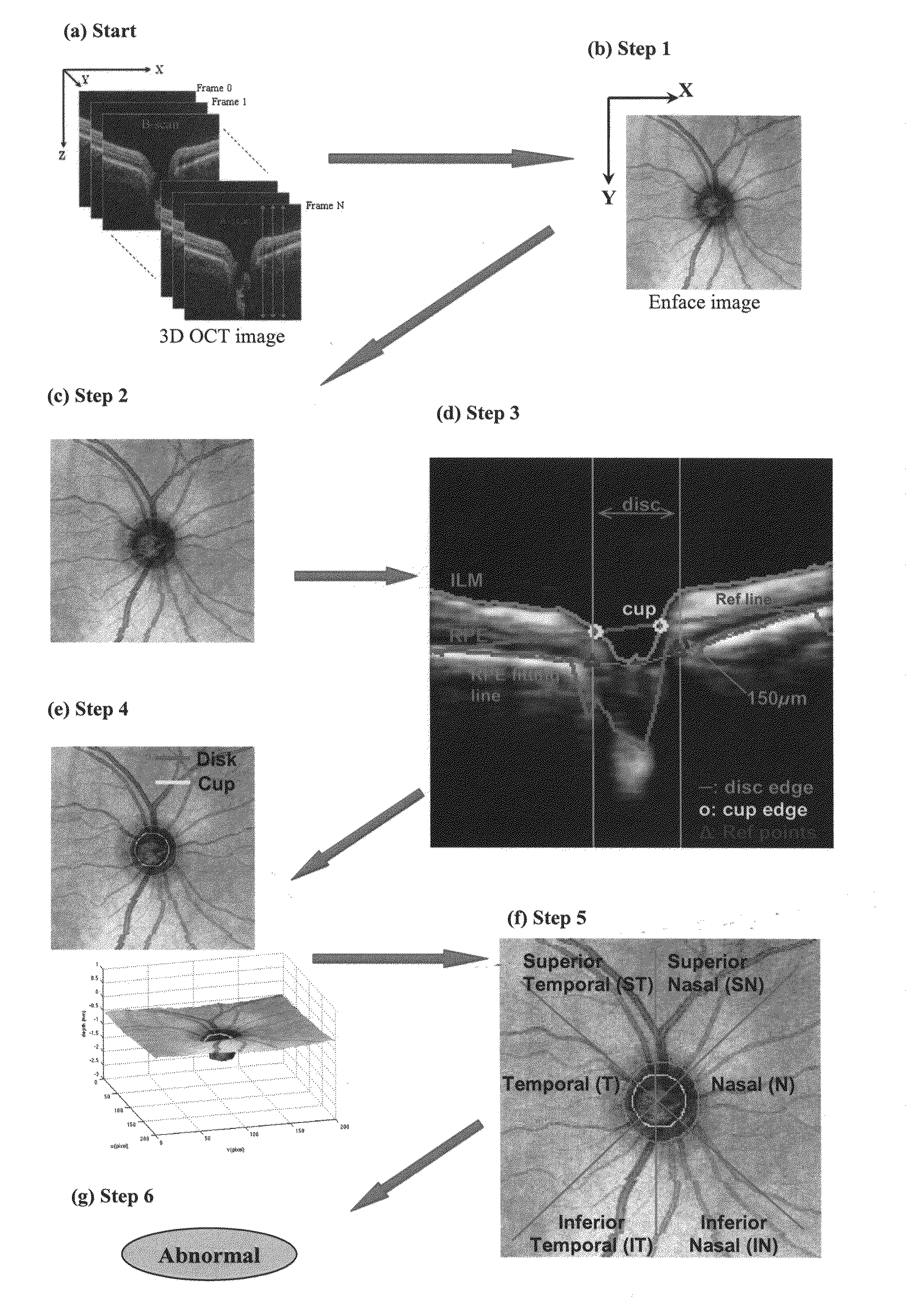
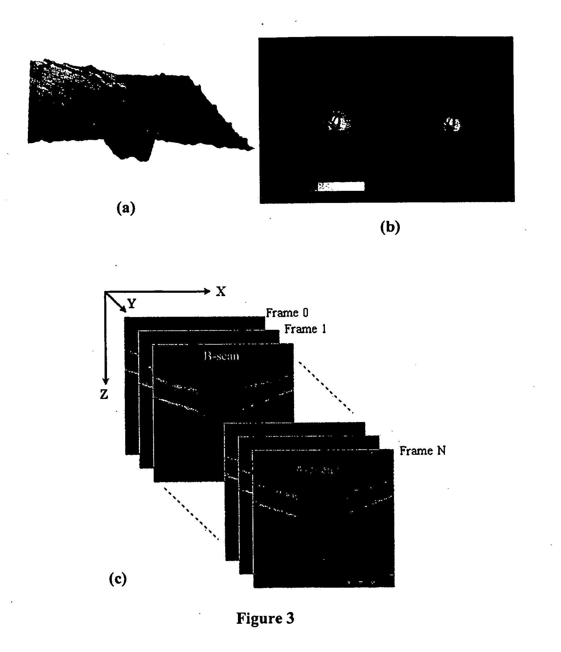
[0041]All the references mentioned herein are incorporated by reference in their entirety. As noted above, the overall system of the present invention is diagramed schematically in FIGS. 6(a)-6(g). A detailed description of the procedures is given as follows:[0042]1. Generate enface image (2D ONH image) from the 3D OCT image taken from SD-OCT, as shown in FIG. 6(a) and / or FIG. 3(c):

[0043](a) The enface image is generated by averaging the intensity values of each A-scan line.

[0044](b) The intensity values is normalized to obtain a high contract enface image.

[0045]Step 1, generation of enface image, comports with existing methodology (FIG. 6(b)). The current SD-OCT machines of several different brands already have implemented enface image generation, based on this basic idea [5]. Data can be utilized from different modes of scanning protocols, such as A-scan, B-scan, and C-scan.[0046]2. Detect the disc margin on 2D ONH image (FIG. 6(c))

[0047]Automated detection of disc margin is a cha...


PUM


Abstract

A fully automated optic nerve head assessment system, based on spectral domain optical coherence tomography, provides essential disc parameters for clinical analysis, early detection, and monitoring of progression.

Description

RELATED PATENT APPLICATIONS[0001]This application claims priority to U.S. Provisional Application Ser. No. 61 / 071,347, filed Apr. 23, 2008, which is incorporated by reference in its entirety.STATEMENT REGARDING FEDERALLY SPONSORED RESEARCH[0002]This invention was made with Government support under grant number RO1-EY013178-8, awarded by the National Institutes of Health. The government has certain rights in the invention.BACKGROUND OF THE INVENTION[0003]Glaucoma is a group of diseases of the eye characterized by progressive damage of the optic nerve that may result in visual field loss and eventual irreversible blindness if inadequately controlled. The World Health Organization currently ranks glaucoma as the second most common cause of blindness. As majority of patients do not experience any symptoms until the disease reaches an advanced stage, early detection and treatment is of utmost importance.[0004]The diagnosis of glaucoma is based on the appearance of the optic nerve head (O...

Claims


Application Information

Patent Timeline
29 Oct 2009
Publication
US20090268159A1
IPC
A61B3/14
CPC
A61B3/102; A61B3/1233; A61B3/14; G06T7/0012; G06T2207/30041; G06T7/402; G06T2207/10016; G06T2207/10072
Inventors
XU, JUAN; WOLLSTEIN, CHAIM-GADI