Imaging table protective cover

a protective cover and imaging table technology, applied in the field of imaging table protective cover, can solve the problems of interference with the operation of the imaging apparatus, contamination of the surface of the support, and the particular vulnerability of the imaging components carried by the patient support to fluids originating from the patient, so as to facilitate cleaning of the cover, facilitate cleaning, and facilitate the effect of patient comfor

Inactive Publication Date: 2006-08-01
DOMICO MED DEVICE LLC
View PDF12 Cites 16 Cited by
  • Summary
  • Abstract
  • Description
  • Claims
  • Application Information

AI Technical Summary

Benefits of technology

[0006]Apparatus constructed in accordance with the invention comprises a sheet formed of pliable, waterproof material which is adapted to overlie the patient supporting surface of a table or other support in which components of imaging apparatus are housed. The sheet has a length corresponding substantially to that of the support surface and a width greater than that of the support surface so that when the sheet is applied to the surface of the table a central section of the sheet rests upon the patient supporting surface of the table and is flanked by flaps which extend beyond and depend from the table surface. Preferably, the center section of the cover includes a pad of cushioning material so as to enhance the comfort of the patient. Preferably, the entire sheet, including the flaps, is encapsulated in a coating of pliable, durable, easily cleanable material so as to facilitate cleaning of the cover between successive uses thereof.
[0009]The materials from which the apparatus is formed are inert insofar as the radiologic imaging apparatus is concerned, thereby avoiding any interference with the imaging procedures.

Problems solved by technology

The imaging components carried by the patient support are particularly vulnerable to fluids originating from a patient or spilled by technicians.
Such fluids have a tendency to collect atop the patient support, or even seep into the support, resulting in contamination of the surface of the support and interference with the operation of the imaging apparatus.
Further, the collection of fluids requires careful cleaning which often consumes so much time that the imaging apparatus cannot be used when needed.
The problems associated with the collection of fluids atop patient supports have been considered heretofore and solutions proposed, but not all of the proposed solutions have been satisfactory for a number of reasons.
For example, some protective covers proposed heretofore are exceedingly complicated in construction, thereby delaying the application and removal of such covers to and from a patient's support.
If such a cover is one which cannot be cleaned and reused, the cost of utilization of such covers is unacceptably high.
In some instances, however, fixed restraints such as straps cannot be used if they overlie or bear against a traumatized portion of the patient's body.
However, not all restraining straps are sufficiently adjustable relative to the patient to provide the necessary restraint at an appropriate position of the patient's body.

Method used


Image

  • Imaging table protective cover
  • Imaging table protective cover
  • Imaging table protective cover

Examples


Embodiment Construction

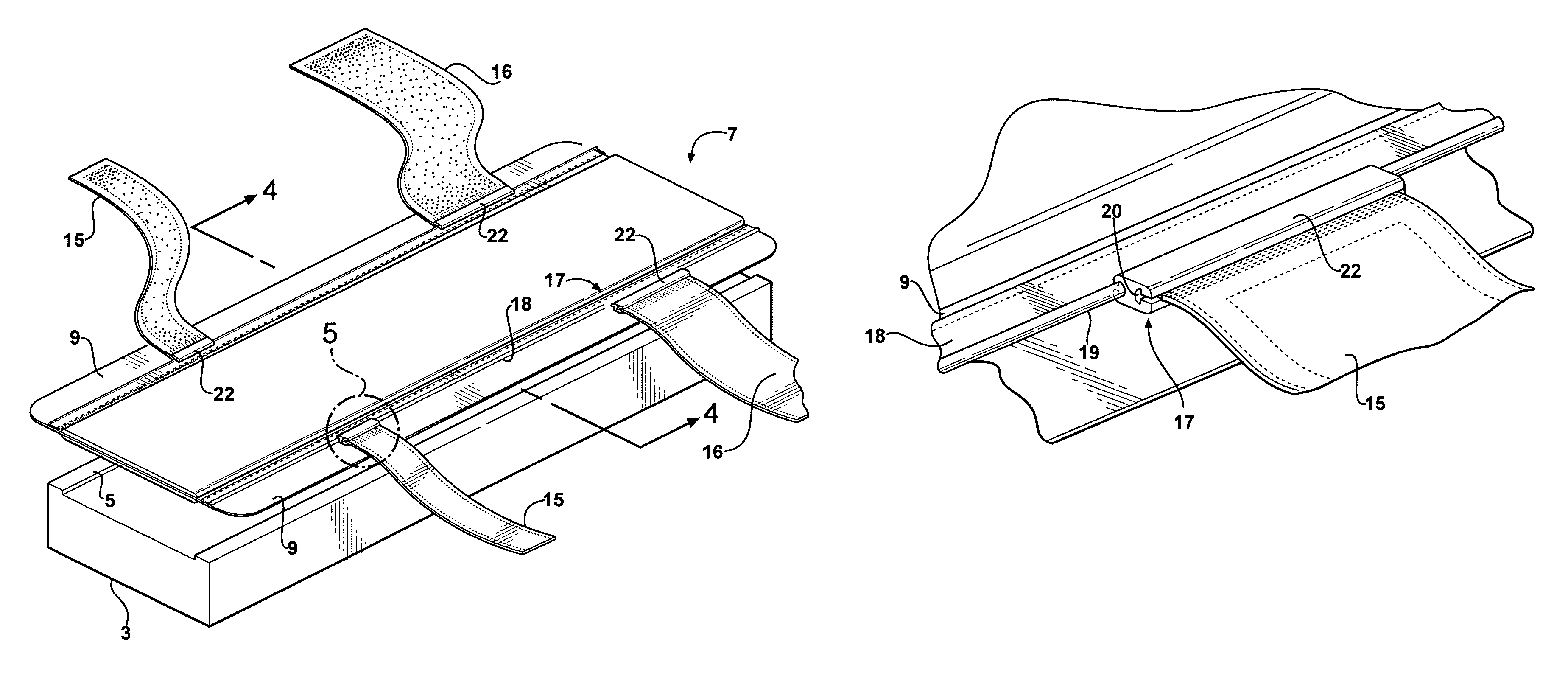
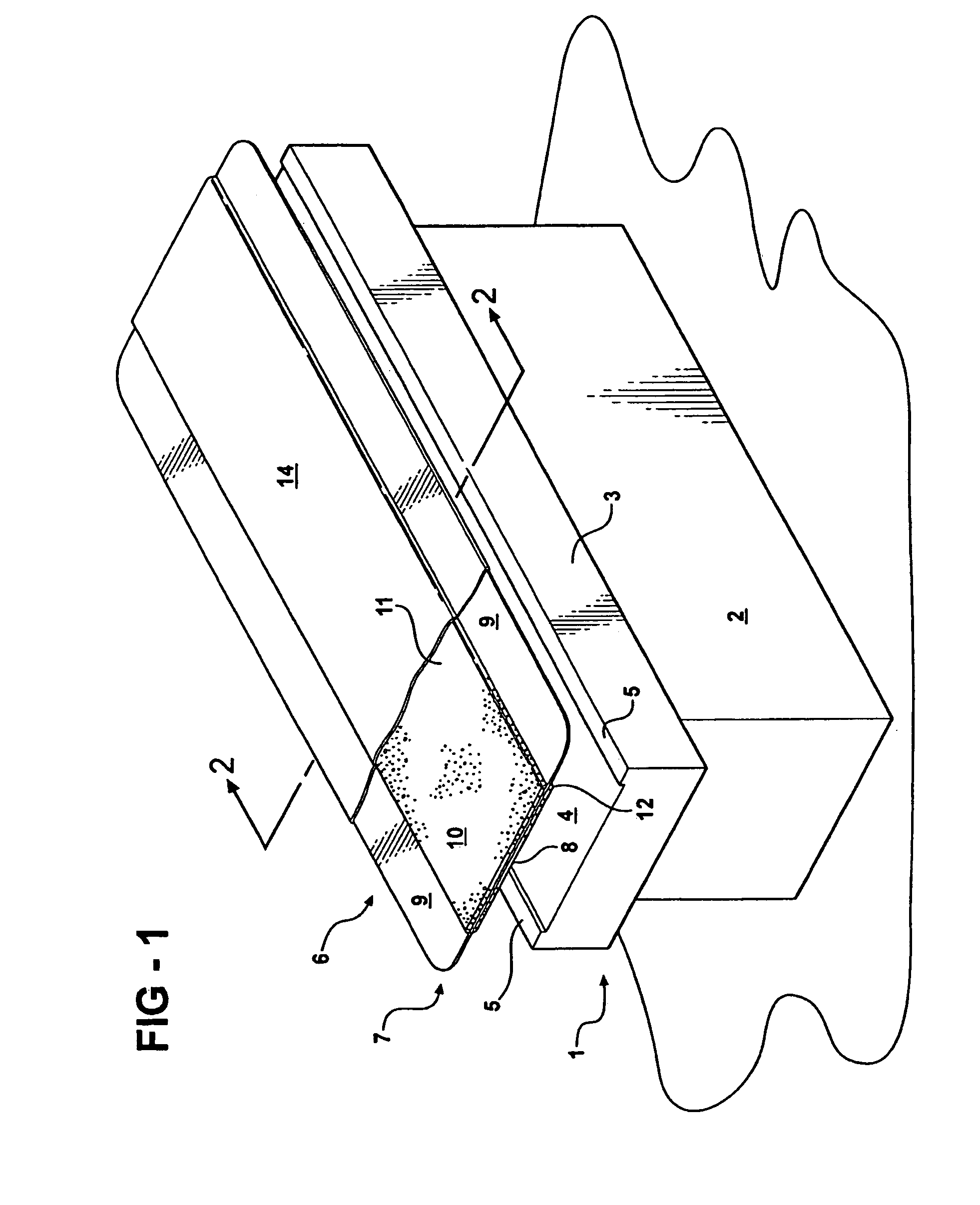
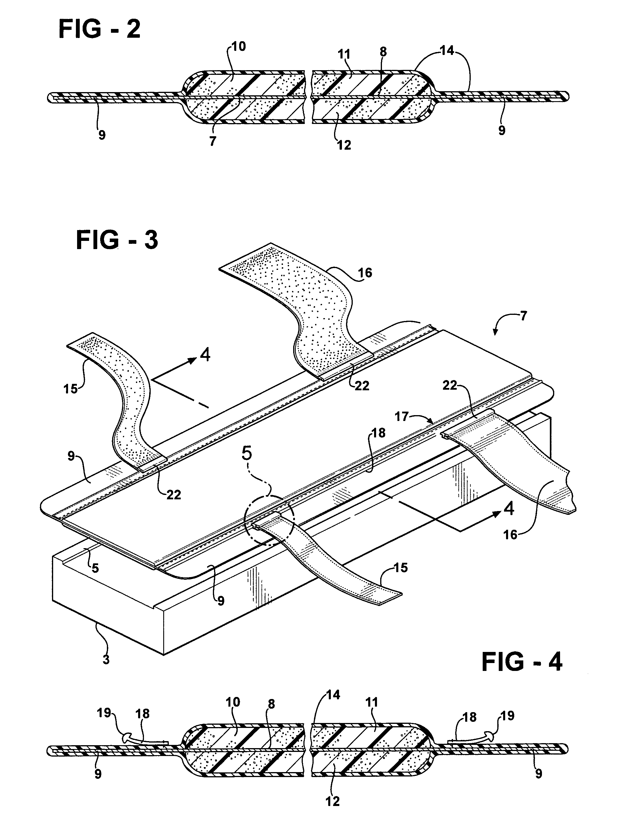
[0019]Apparatus constructed in accordance with the invention is adapted for use with a support table 1 having a base 2 supporting a top 3 having an upper surface 4 flanked by longitudinally extending rails 5. The base 2, or the top 3, or both, include radiologic imaging components (not shown) which cooperate with other components contained in an overhead support (not shown) for producing diagnostic images of a patient lying atop the table. The rails 5 help define the area to be occupied by the patient during the diagnostic process. However, the rails, if used, tend to enable fluids to collect on the upper surface of the table, thereby making cleaning of the table between uses more complicated and, perhaps, enabling such fluids to seep into the interior of the support.

[0020]The apparatus comprises a cover 6 formed from a sheet 7 of waterproof, pliable material such as polyvinyl chloride which is inert with respect to the imaging apparatus. The sheet 7 has a center section 8 having a ...


PUM


Abstract

A cover and patient restraint for use with a radiologic imaging table having an upper surface on which a patient may lie, the cover having a central section corresponding generally to the length and width of the patient supporting surface of the table and a width substantially greater than that of the supporting surface so the opposite sides of the cover form flaps which extend beyond and depend from the upper surface of the table to prevent the collection on the table surface of liquids. The cover includes a pad of cushioning material inboard of the flaps to enhance the comfort of a patient lying on the cover. Restraining straps are coupled to anchor strips extending longitudinally of the cover adjacent opposite sides of the pad so as to enable the straps to be placed in overlying relation to the patient in any selected one of a large number of adjusted positions. The cover is encapsulated in a pliable, washable, radiologic inert coating.

Description

[0001]This invention relates to a protective cover for use in connection with a radiologic imaging table.BACKGROUND OF THE INVENTION[0002]Radiologic imaging is used extensively for diagnostic procedures and the monitoring of patients. The imaging apparatus conventionally includes a table or other support on which a patient may lie for exposure to imaging apparatus having cooperable components positioned below and above the patient. The imaging components carried by the patient support are particularly vulnerable to fluids originating from a patient or spilled by technicians. Such fluids have a tendency to collect atop the patient support, or even seep into the support, resulting in contamination of the surface of the support and interference with the operation of the imaging apparatus. Further, the collection of fluids requires careful cleaning which often consumes so much time that the imaging apparatus cannot be used when needed.[0003]The problems associated with the collection of...

Claims


Application Information

Patent Timeline
Patent Type & Authority Patents(United States)
IPC IPC(8): A47G9/04
CPCA61B6/0421A61G13/102A61G2210/50
Inventor CZOP, MICHAEL W.
Owner DOMICO MED DEVICE LLC
Why Eureka
Try Eureka