
Electrokinetic concentration device and methods of use thereof
a technology of electrokinetic concentration and concentration device, which is applied in the field of electrokinetic concentration device, can solve the problems of sample volume mismatch, inability to meet the requirements of sample volume, so as to reduce or enhance the adsorption of said species, enhance or reduce the operation efficiency of the device, enhance or reduce the effect of the devi
- Summary
- Abstract
- Description
- Claims
- Application Information
AI Technical Summary
Benefits of technology
Problems solved by technology
Method used
Image

Examples
example 1
Concentration of Charged Species Via Electroosmosis and Electrokinetic Trapping
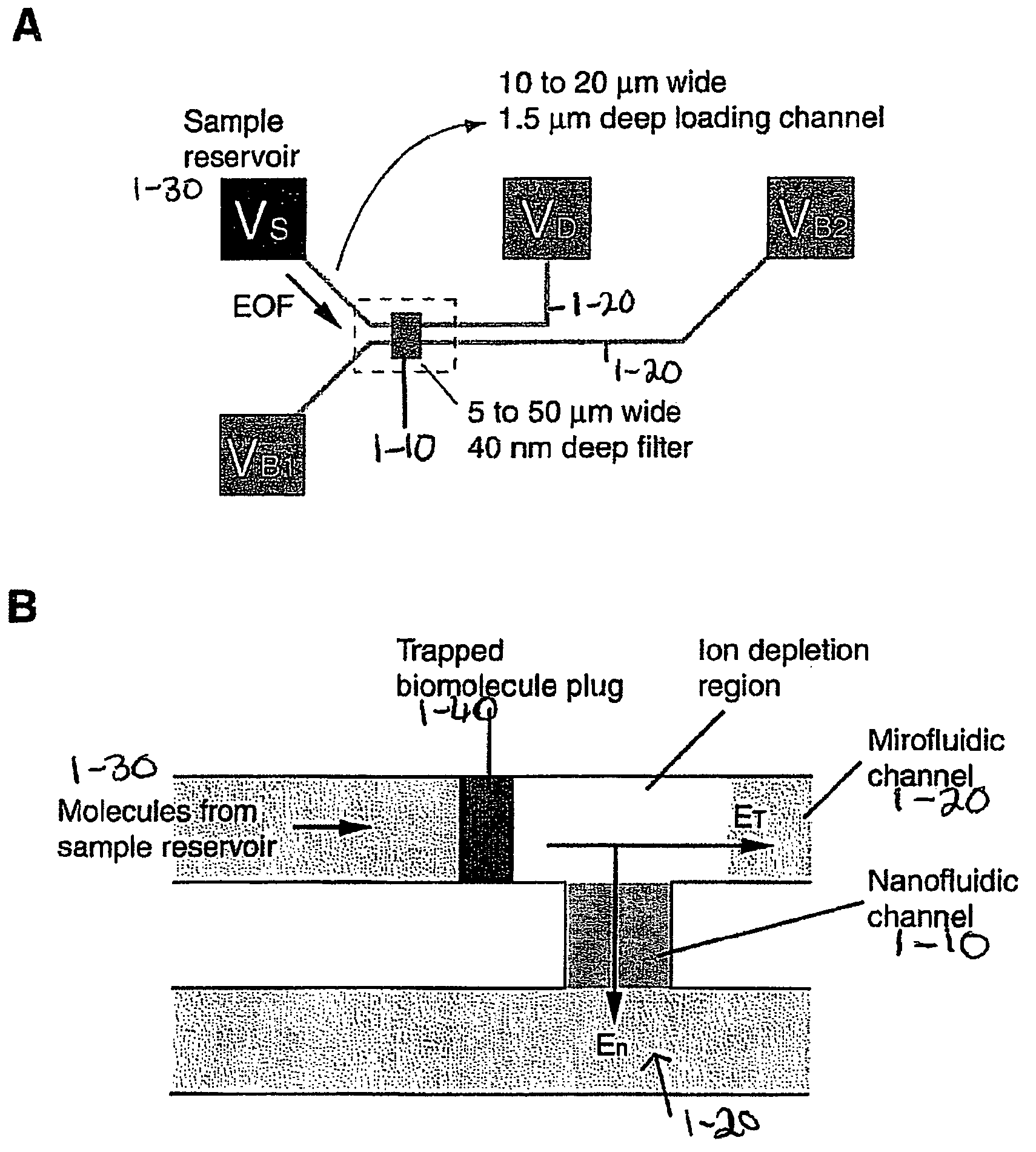
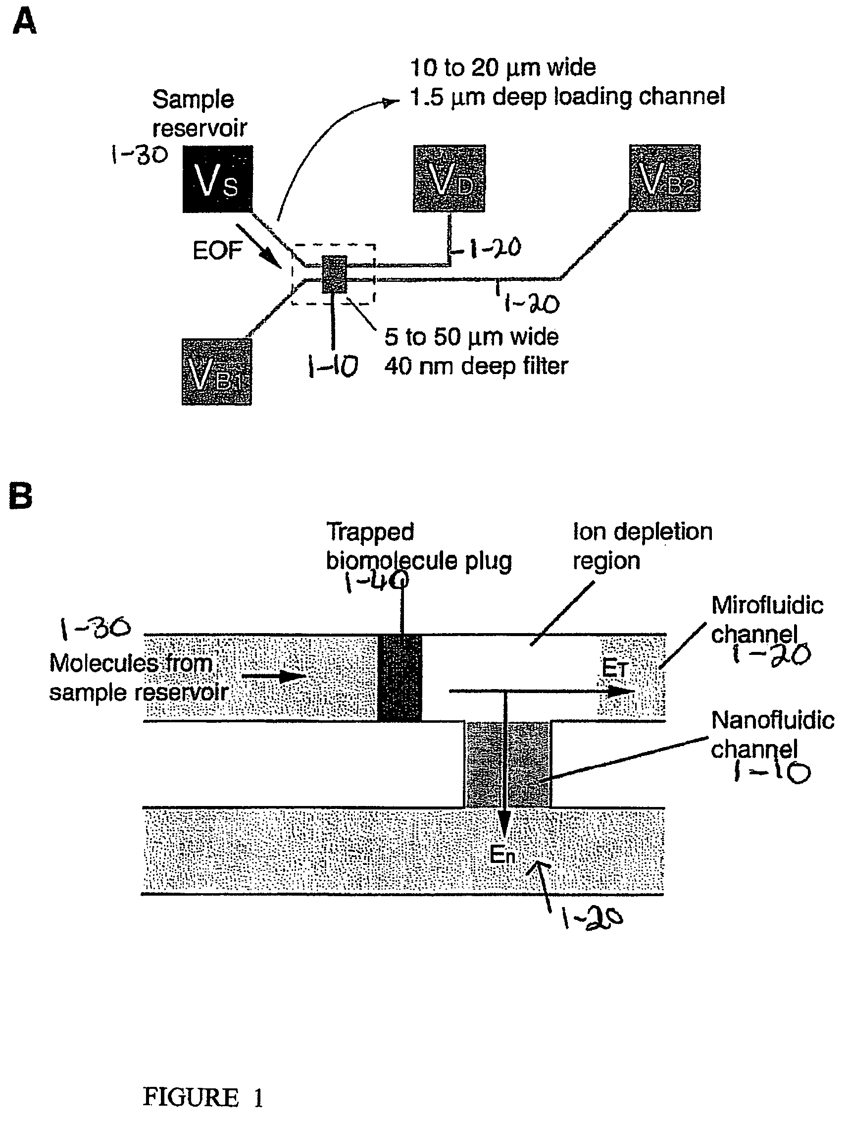
[0147]Biomolecules are efficiently concentrated in a device, which provides for a nonlinear electroosmotic flow (much stronger than normal electroosmotic flow) generated in a channel of the device, conveying biomolecules to a “trapping region” with a high flow speed, in combination with an energy barrier for the molecules generated by an induced space charge layer in the device. The combination of these two phenomena results in a rapid concentration of charged biomolecules, for example, proteins and peptides within a microfluidic channel of the device, without any physical barriers or reagents, up to 106˜108 fold in concentration.
[0148]One embodiment of the device is schematically depicted in FIGS. 1A and B. One or many thin nanofluidic channels (1-10), typically less than 50 nm in thickness, connect at least two microfluidic channels (1-20). Two separate electric fields are applied to the device, and con...
example 2
Protein Concentration Via Electroosmosis and Electrokinetic Trapping
[0156]FIG. 4 demonstrates a procedure used for the quantification of the molecular concentration in the channel. The embodied device can generate sample plugs that saturate the CCD array used for detection, and thus neutral density filters may be used. The concentration of collected GFP protein plugs was estimated by measuring the fluorescence signal from the molecules in the channels. Channels filled with 3.3 μM and 0.33 μM GFP solutions, allowed for little detection of the 0.33 μM GFP solution (FIG. 4C). The two intensity levels were compared with local intensity level after preconcentration of the molecules (FIG. 4D).
[0157]FIG. 5 demonstrates the stability and performance of the embodied device. In FIG. 5(A-C), a solution of 33 M GFP (green fluorescent protein) was loaded into the sample reservoir, and the resulting trapped protein peaks were monitored by fluorescence microscopy. Protein concentration was stably ...
example 3
Analysis of Proteins Concentrated Via Electroosmosis and Electrokinetic Trapping
[0164]Molecular trapping in the embodied device can be turned off by removing En. The buffer solution is expected to re-establish the ionic balance as soon as the field is turned off. When the field is turned off, the collected peak is instantly dispersed to about twice the original peak width, but there is no further dispersion observed. Therefore, the molecular plug generated by the embodied device can be easily manipulated either by electric field (electrophoresis) or pressure-driven flow. FIG. 6 demonstrates capillary electrophoresis of two protein species collected by the embodied device. The results indicate effective coupling of the embodied device and downstream analysis tools, in this case, free solution capillary electrophoresis.
PUM
| Property | Measurement | Unit |
|---|---|---|
| voltage | aaaaa | aaaaa |
| voltage | aaaaa | aaaaa |
| width | aaaaa | aaaaa |
Abstract
Description
Claims
Application Information