Electrokinetic concentration device and methods of use thereof

a technology of electrokinetic concentration and concentration device, which is applied in the field of electrokinetic concentration device, can solve the problems of sample volume mismatch, inability to meet the requirements of sample volume, so as to reduce or enhance the adsorption of said species, enhance or reduce the operation efficiency of the device, enhance or reduce the effect of the devi

Inactive Publication Date: 2006-08-17
MASSACHUSETTS INST OF TECH
View PDF5 Cites 74 Cited by
  • Summary
  • Abstract
  • Description
  • Claims
  • Application Information

AI Technical Summary

Benefits of technology

[0015] In one embodiment, the surface of the microchannel has been functionalized to reduce or enhance adsorption of said species of interest to said surface. In another embodiment, the surface of the nanochannel and / or microchannel has been functionalized to enhance or reduce the operation efficiency of the device. In another embodiment, external gate potential is applied to the substrate of the device, to enhance or reduce the operation efficiency of the device. In another embodiment, the device is comprised of a transparent material. In another embodiment, the transparent material is pyrex, silicon dioxide, silicon nitride, quartz or SU-8.

Problems solved by technology

One of the major challenges of proteomics is the sheer complexity of biomolecule samples, such as blood serum or cell extract.
Such diversity of proteins, as well as their huge concentration ranges, poses a formidable challenge for sample preparation in proteomics.
Conventional protein analysis techniques, based on multidimensional separation steps and mass spectrometry (MS), fall short because of the limited separation peak capacity (up to ˜3000) and dynamic range of detection (˜104).
However, most microfluidic separation and sample processing devices suffers from the critical issue of sample volume mismatch.
Therefore, microchip-based separation techniques often analyze only a small fraction of available samples, which significantly limits the overall detection sensitivity.
In proteomics, this problem is exacerbated by the fact that information-rich signaling molecules (cytokines and biomarkers, e.g.) are present only in trace concentrations (nM˜pM range), and there is no signal amplification technique such as polymerase chain reaction (PCR) for proteins and peptides.
Overall, the demonstrated concentration factors for the existing preconcentration schemes are limited to ˜1000, and their application to the integrated Microsystems is difficult due to various operational constraints such as reagents and materials requirements.

Method used


Image

  • Electrokinetic concentration device and methods of use thereof
  • Electrokinetic concentration device and methods of use thereof
  • Electrokinetic concentration device and methods of use thereof

Examples


example 1

Concentration of Charged Species Via Electroosmosis and Electrokinetic Trapping

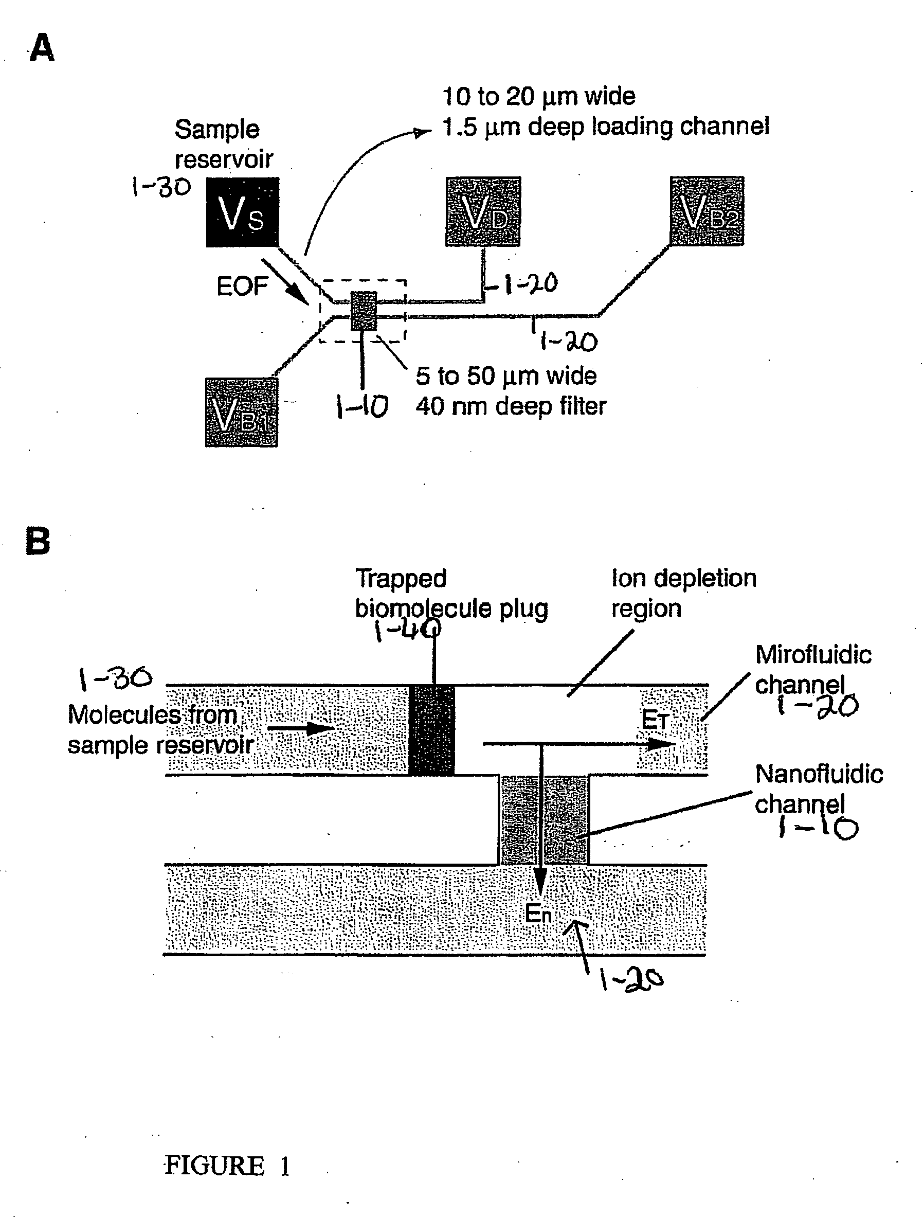
[0150] Biomolecules are efficiently concentrated in a device, which provides for a nonlinear electroosmotic flow (much stronger than normal electroosmotic flow) generated in a channel of the device, conveying biomolecules to a “trapping region” with a high flow speed, in combination with an energy barrier for the molecules generated by an induced space charge layer in the device. The combination of these two phenomena results in a rapid concentration of charged biomolecules, for example, proteins and peptides within a microfluidic channel of the device, without any physical barriers or reagents, up to 106˜108 fold in concentration.

[0151] One embodiment of the device is schematically depicted in FIGS. 1A and B. One or many thin nanofluidic channels (1-10), typically less than 50 nm in thickness, connect at least two microfluidic channels (1-20). Two separate electric fields are applied to the device, and...

example 2

Protein Concentration Via Electroosmosis and Electrokinetic Trapping

[0159]FIG. 4 demonstrates a procedure used for the quantification of the molecular concentration in the channel. The embodied device can generate sample plugs that saturate the CCD array used for detection, and thus neutral density filters may be used. The concentration of collected GFP protein plugs was estimated by measuring the fluorescence signal from the molecules in the channels. Channels filled with 3.3 μM and 0.33 μM GFP solutions, allowed for little detection of the 0.33 μM GFP solution (FIG. 4C). The two intensity levels were compared with local intensity level after preconcentration of the molecules (FIG. 4D).

[0160]FIG. 5 demonstrates the stability and performance of the embodied device. In FIG. 5(A-C), a solution of 33 μM GFP (green fluorescent protein) was loaded into the sample reservoir, and the resulting trapped protein peaks were monitored by fluorescence microscopy. Protein concentration was stab...

example 3

Analysis of Proteins Concentrated Via Electroosmosis and Electrokinetic Trapping

[0167] Molecular trapping in the embodied device can be turned off by removing En. The buffer solution is expected to re-establish the ionic balance as soon as the field is turned off. When the field is turned off, the collected peak is instantly dispersed to about twice the original peak width, but there is no further dispersion observed. Therefore, the molecular plug generated by the embodied device can be easily manipulated either by electric field (electrophoresis) or pressure-driven flow. FIG. 6 demonstrates capillary electrophoresis of two protein species collected by the embodied device. The results indicate effective coupling of the embodied device and downstream analysis tools, in this case, free solution capillary electrophoresis.


PUM

PropertyMeasurementUnit
voltageaaaaaaaaaa
voltageaaaaaaaaaa
widthaaaaaaaaaa

Abstract

The present invention provides a device and methods of use thereof in concentrating a species of interest and / or controlling liquid flow in a device. The methods, inter-alia, make use of a device comprising microchannels, which are linked to nanochannels, whereby induction of an electric field in the nanochannel results in ion depletion in the linkage region between the microchannel and nanochannel, and a space charge layer is formed within the microchannel, which provides an energy barrier for said species of interest which enables its concentration in a region in the microchannel.

Description

CROSS REFERENCE TO RELATED APPLICATIONS [0001] This invention claims the benefit of U.S. Provisional Application Ser. No. 60 / 646,513, which was filed Jan. 25, 2005, which is hereby incorporated in its entirety.FIELD OF THE INVENTION [0002] This invention provides devices and methods of use thereof in concentrating a charged species of interest in solution. This invention provides a concentration device, which is based on electrokinetic trapping of a charged species of interest, which can be further isolated and analyzed. BACKGROUND OF THE INVENTION [0003] One of the major challenges of proteomics is the sheer complexity of biomolecule samples, such as blood serum or cell extract. Typical blood samples could contain more than 10,000 different protein species, with concentrations varying over 9 orders of magnitude. Such diversity of proteins, as well as their huge concentration ranges, poses a formidable challenge for sample preparation in proteomics. [0004] Conventional protein analy...

Claims


Application Information

Patent Timeline
Patent Type & Authority Applications(United States)
IPC IPC(8): G01N27/00
CPCB01L3/502753G01N27/44791Y10S977/904Y10S977/924B81B7/00G01N27/00G01N33/00
Inventor HAN, JONGYOONWANG, YING-CHIH
Owner MASSACHUSETTS INST OF TECH
Why Eureka
Try Eureka