Method and system for diaphragm segmentation in chest X-ray radiographs

a radiograph and diaphragm technology, applied in the field of diaphragm segmentation in a chest x-ray radiograph, can solve the problems of not much edge information and/or intensity information that provides sufficient, and the problem of poorly addressed diaphragm segmentation

Active Publication Date: 2011-12-06
SIEMENS HEALTHCARE GMBH
View PDF5 Cites 0 Cited by
  • Summary
  • Abstract
  • Description
  • Claims
  • Application Information

AI Technical Summary

Problems solved by technology

However, even though many techniques have been developed for segmentation of lung lobes, heart regions, and ribs in chest x-ray radiographs, diaphragm segmentation has not been well addressed.
Furthermore, diaphragm regions typically do not have a clear-cut definition in chest x-ray images, and there is not much edge information and / or intensity information that provides sufficient support segmenting diaphragm regions using a general segmentation algorithm.

Method used


Image

  • Method and system for diaphragm segmentation in chest X-ray radiographs
  • Method and system for diaphragm segmentation in chest X-ray radiographs
  • Method and system for diaphragm segmentation in chest X-ray radiographs

Examples


Embodiment Construction

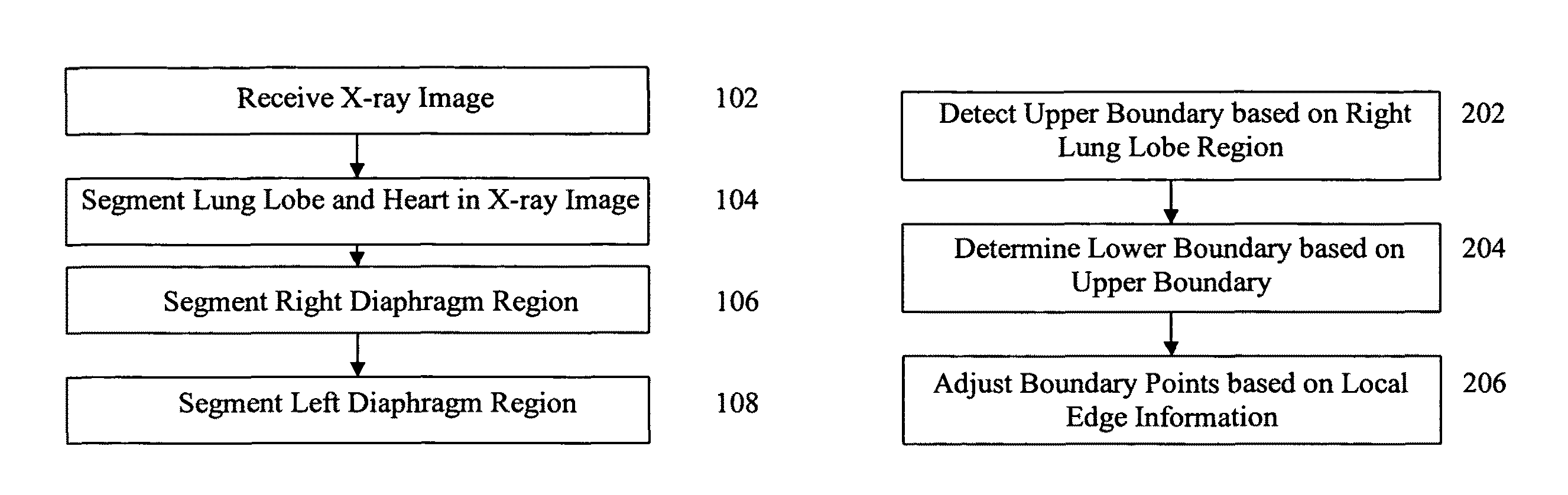
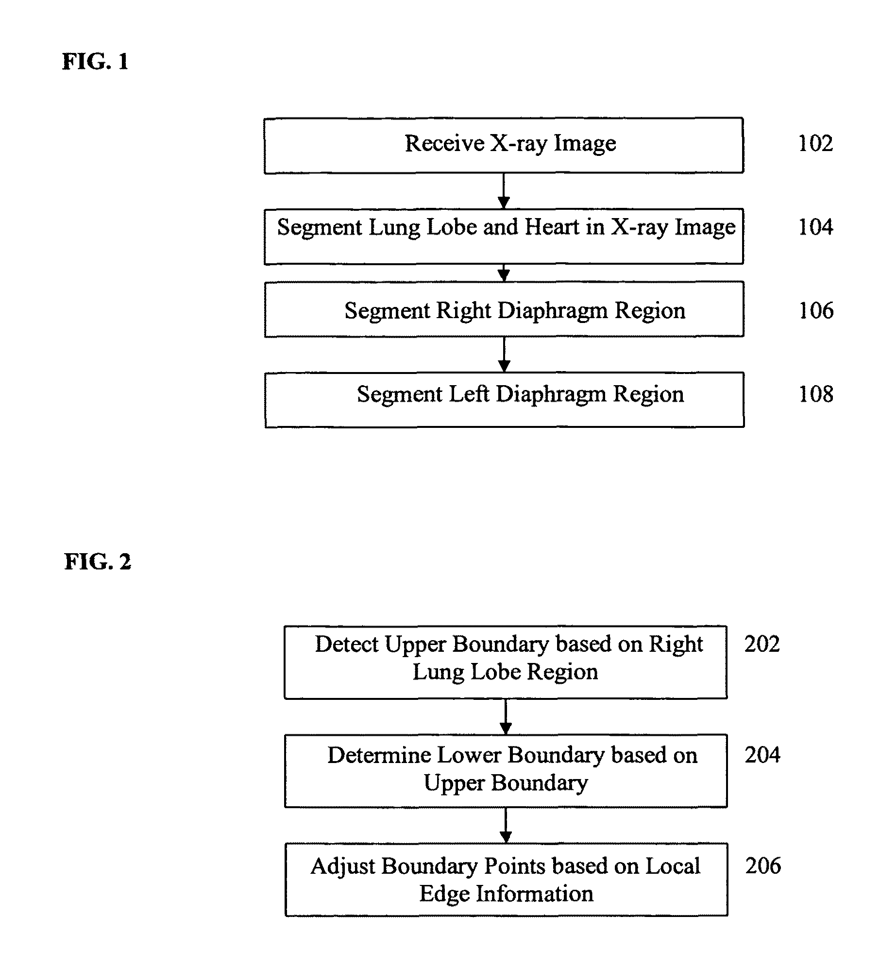
The present invention is directed to a method for segmenting diaphragm regions in an X-ray radiograph. Embodiments of the present invention are described herein to give a visual understanding of the segmentation method. A digital image is often composed of digital representations of one or more objects (or shapes). The digital representation of an object is often described herein in terms of identifying and manipulating the objects. Such manipulations are virtual manipulations accomplished in the memory or other circuitry / hardware of a computer system. Accordingly, is to be understood that embodiments of the present invention may be performed within a computer system using data stored within the computer system.

Embodiments of the present invention are directed to segmenting diaphragm regions in a chest X-ray radiograph. The segmentation of the diaphragm regions, according to embodiments of the present invention, is based in part on boundaries of the lungs acquired using a lung segme...


PUM


Abstract

A method and system for segmenting diaphragm regions in a chest X-ray radiograph is disclosed. The diaphragm regions are segmented based on left and right lung lobe regions and a heart region in the chest X-ray radiograph. A right diaphragm region is segmented in the chest X-ray radiograph based a boundary of the right lung lobe. A left diaphragm region is segmented in the chest X-ray radiograph based on the heart region and a boundary of the left lung lobe.

Description

BACKGROUND OF THE INVENTIONThe present invention relates to diaphragm segmentation in a chest X-ray radiograph, and more particularly, to estimating regions representing the diaphragm in a chest X-ray radiograph, for nodule detection in the diaphragm region.Nodules are small masses of tissue, which can form on various organs in the human body, such as the lungs. Lung nodule detection can be used for advance cancer screening. Accordingly, computer implemented methods for automatic nodule detection methods have been developed to detect nodules in chest X-ray radiographs. Such automatic nodule detection methods determine locations in a chest X-ray radiograph at which there is a high probability that a nodule exists.In chest X-ray radiographs, diaphragm regions typically exhibit unique intensity characteristics. Diaphragm regions can represent special regions of interest for nodule detection in chest X-ray radiographs. For example, in lung nodule detection, detectable nodules in diaphra...

Claims


Application Information

Patent Timeline
Patent Type & Authority Patents(United States)
IPC IPC(8): G06K9/00
CPCG06T7/0083G06T7/0089G06T2207/10116G06T2207/20012G06T2207/20036G06T2207/30061G06T7/12G06T7/149
Inventor HONG, LINLI, YINGSHEN, HONG
Owner SIEMENS HEALTHCARE GMBH
Why Eureka
Try Eureka